

FULL TITLE:
The HCP 7T Retinotopy Dataset: Description and pRF Analysis
SPECIES:
Human
DESCRIPTION:
About a quarter of human cerebral cortex is dedicated mainly to visual processing. The large-scale organization of visual cortex can be measured with functional magnetic resonance imaging (fMRI) while subjects view spatially modulated visual stimuli, also known as 'retinotopic mapping'. One of the datasets collected by the Human Connectome Project (HCP) involved ultra-high-field (7 Tesla) fMRI retinotopic mapping in 181 healthy young adults (1.6-mm resolution), yielding the largest freely available collection of retinotopy data. Here, we describe the experimental paradigm and the results of model-based analysis of the fMRI data. These results provide estimates of population receptive field position and size. Our analyses include both results from individual subjects as well as results obtained by averaging fMRI time-series across subjects at each cortical and subcortical location and then fitting models. Both the group-average and individual-subject results reveal robust signals across much of the brain, including occipital, temporal, parietal, and frontal cortex as well as subcortical areas. The group-average results agree well with previously published parcellations of visual areas. In addition, split-half analyses show strong within-subject reliability, further demonstrating the high quality of the data. We make publicly available the analysis results for individual subjects and the group average, as well as associated stimuli and analysis code. These resources provide an opportunity for studying fine-scale individual variability in cortical and subcortical organization and the properties of high-resolution fMRI. In addition, they provide a set of observations that can be compared with other HCP measures acquired in these same participants.
ABSTRACT:
About a quarter of human cerebral cortex is dedicated mainly to visual processing. The large-scale organization of visual cortex can be measured with functional magnetic resonance imaging (fMRI) while subjects view spatially modulated visual stimuli, also known as 'retinotopic mapping'. One of the datasets collected by the Human Connectome Project (HCP) involved ultra-high-field (7 Tesla) fMRI retinotopic mapping in 181 healthy young adults (1.6-mm resolution), yielding the largest freely available collection of retinotopy data. Here, we describe the experimental paradigm and the results of model-based analysis of the fMRI data. These results provide estimates of population receptive field position and size. Our analyses include both results from individual subjects as well as results obtained by averaging fMRI time-series across subjects at each cortical and subcortical location and then fitting models. Both the group-average and individual-subject results reveal robust signals across much of the brain, including occipital, temporal, parietal, and frontal cortex as well as subcortical areas. The group-average results agree well with previously published parcellations of visual areas. In addition, split-half analyses show strong within-subject reliability, further demonstrating the high quality of the data. We make publicly available the analysis results for individual subjects and the group average, as well as associated stimuli and analysis code. These resources provide an opportunity for studying fine-scale individual variability in cortical and subcortical organization and the properties of high-resolution fMRI. In addition, they provide a set of observations that can be compared with other HCP measures acquired in these same participants.
PUBLICATION:
BioRxiv
- DOI:
10.1101/308247
- Noah Benson
- Keith W. Jamison
- Michael J. Arcaro
- An Vu
- Matthew F. Glasser
- Timothy S. Coalson
- David C. Van Essen
- Essa Yacoub
- Kamil Ugurbil
- Jonathan Winawer
- Kendrick Kay
- University of Minnesota
- Washington University in St. Louis
- New York University
- Harvard University
- St. Luke's Hospital
-
Retinotopy_HCP_7T_181_Fit1.scene
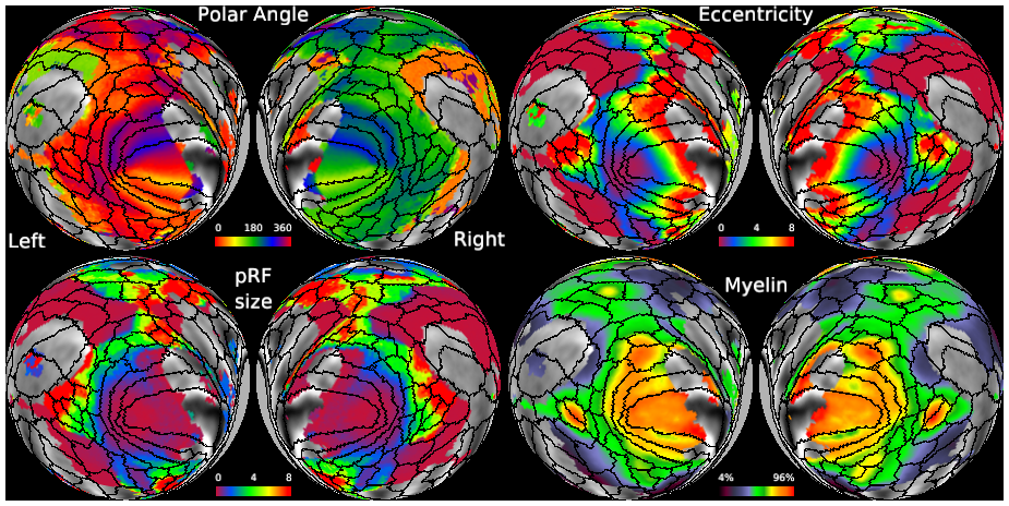
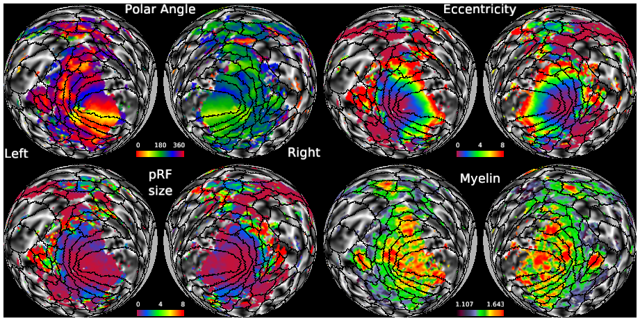
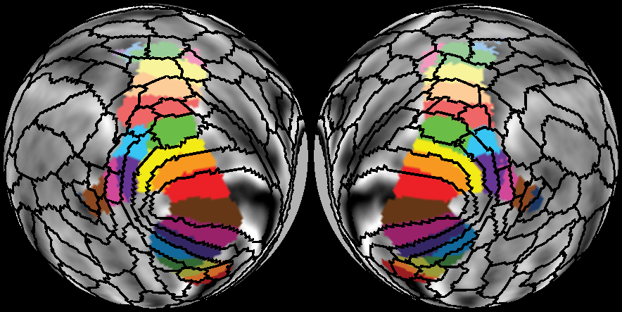
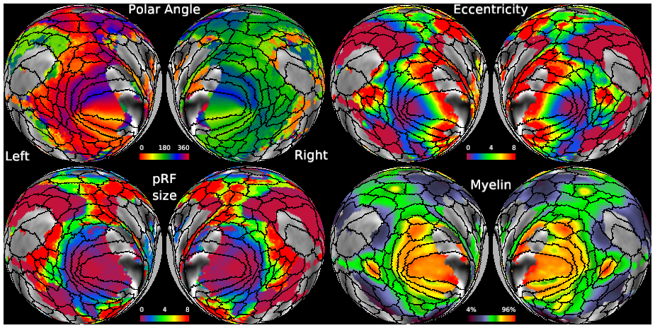
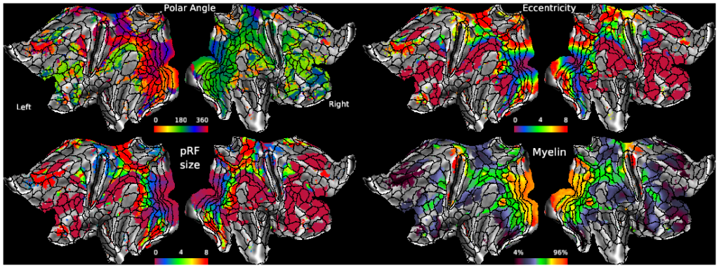
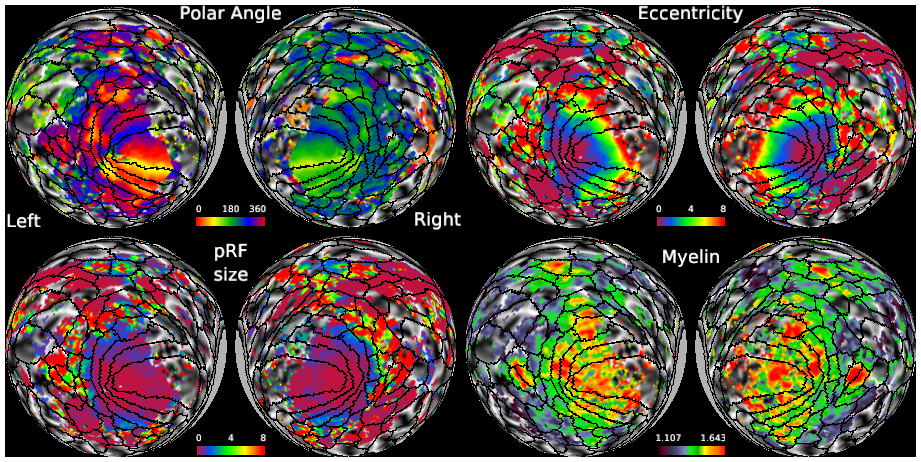
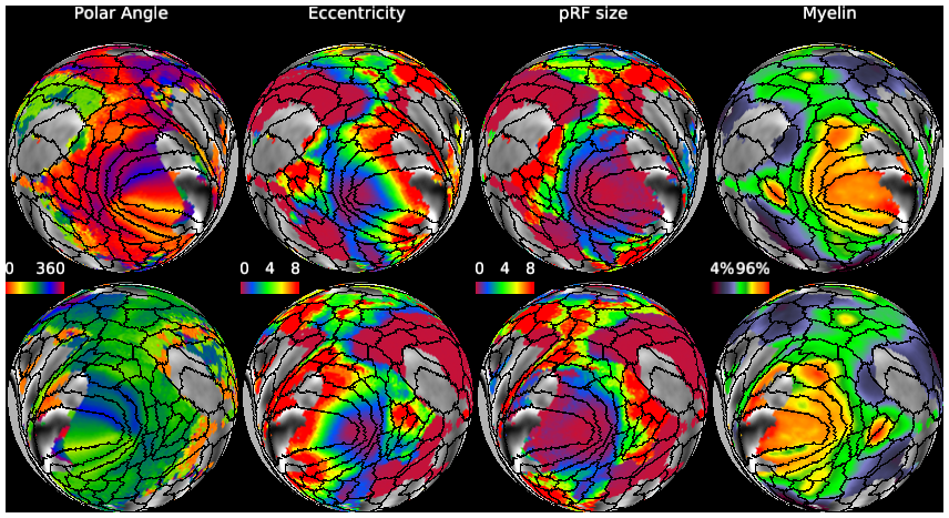
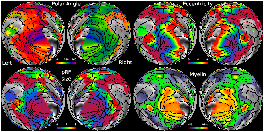
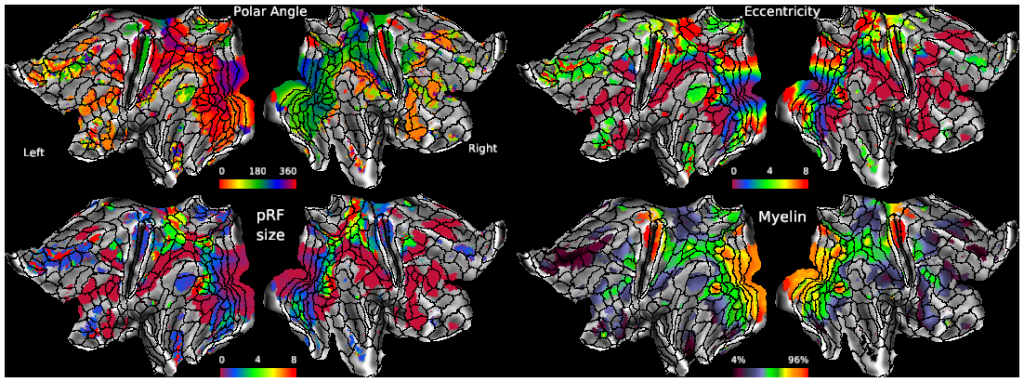
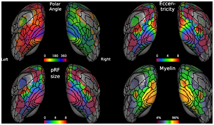
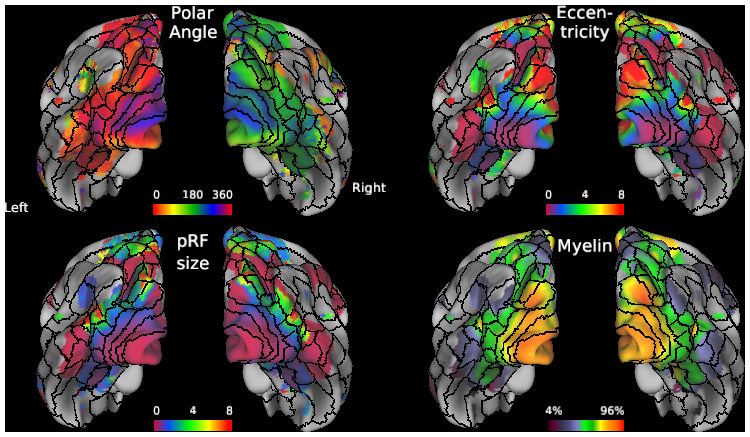
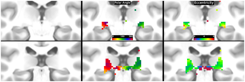
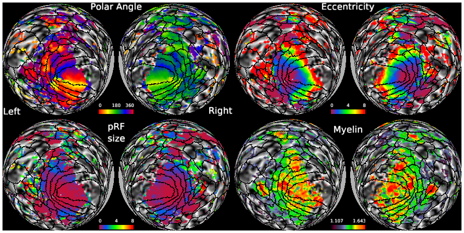
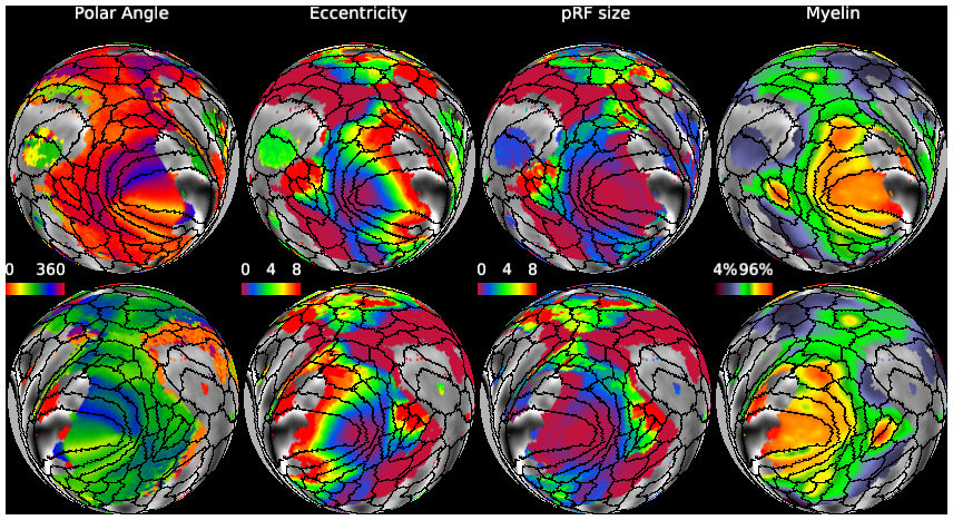
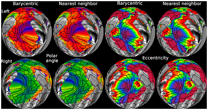
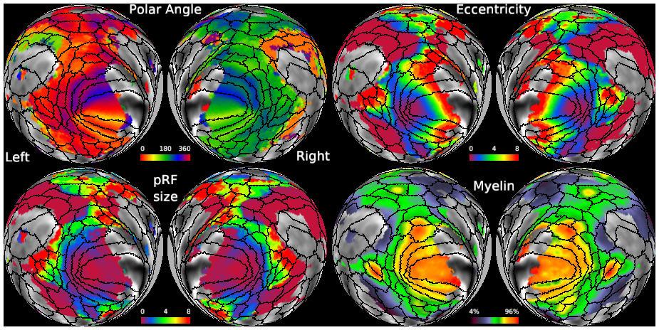
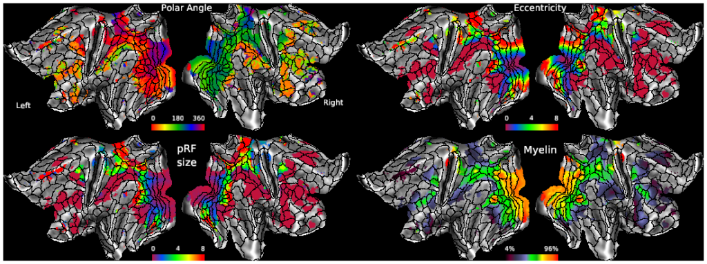
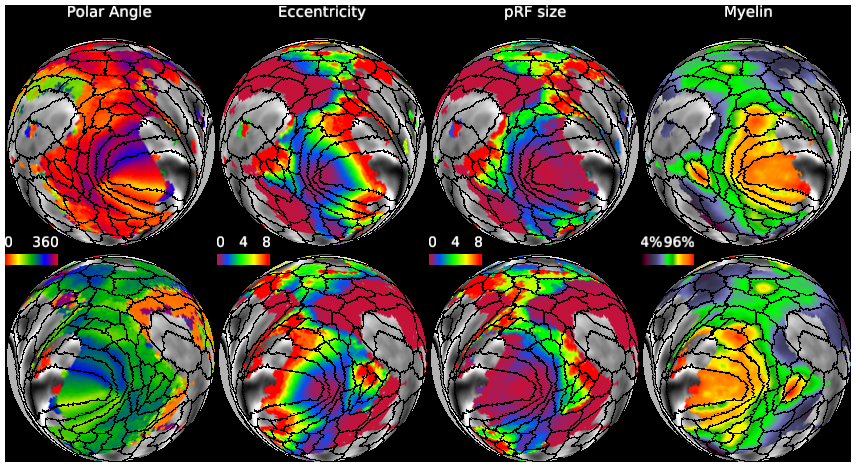
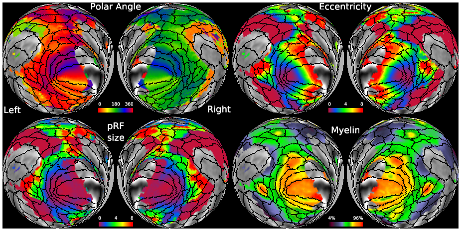
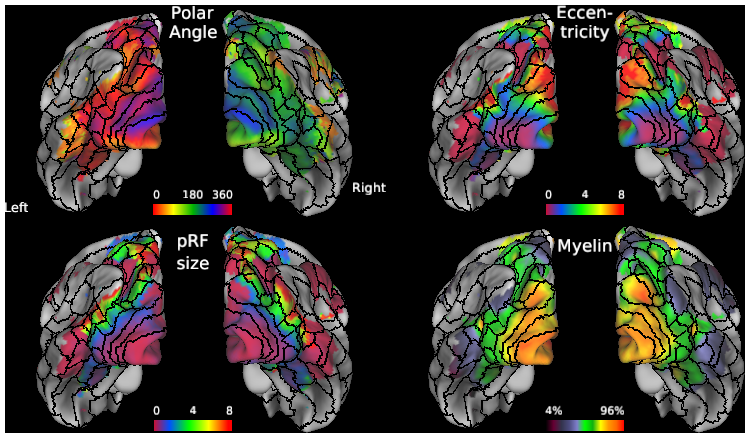
SCENES:- Spheres: Group avg Angle (0 - 360), Eccentricity, pRF, Myelin
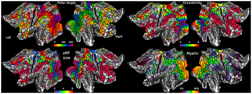
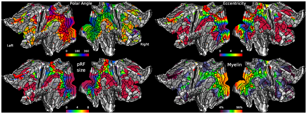
- Flatmaps: Group Avg Angle, Eccentricity, pRF, Myelin
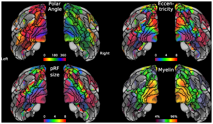
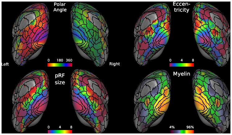
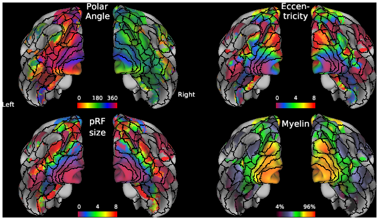
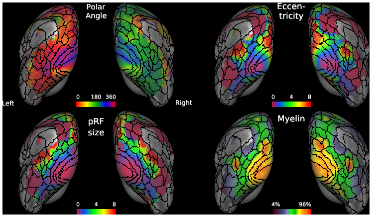
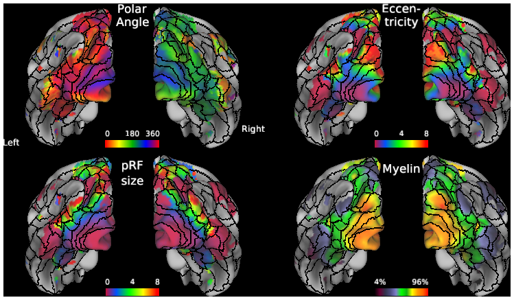
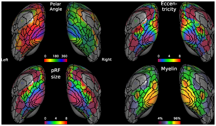
- Inflated: Group Avg Angle, Eccentricity, pRF, Myelin
- Midthickness: Group Avg Angle, Eccentricity, pRF, Myelin
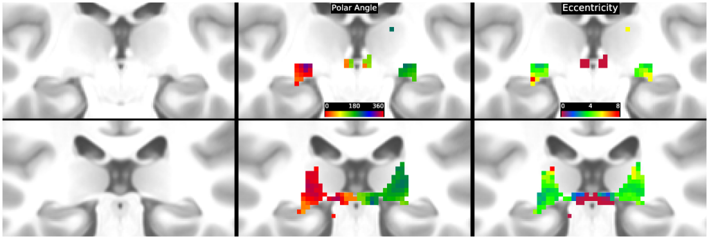
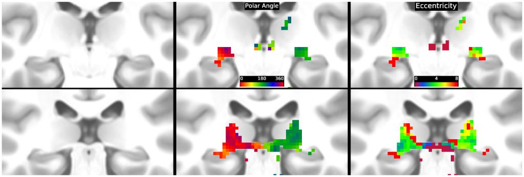
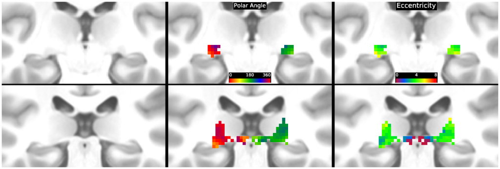
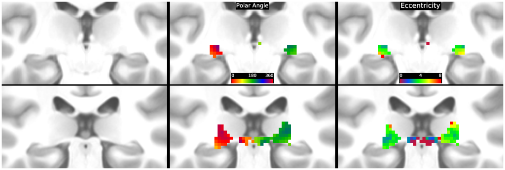
- Subcortical retinotopy: LGN top row); Sup Colliculus (bottom row)
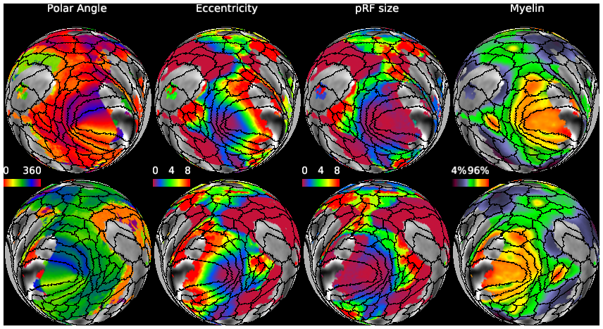
- Spheres: Individual-subject polar angle, eccentricity, pRF, myelin
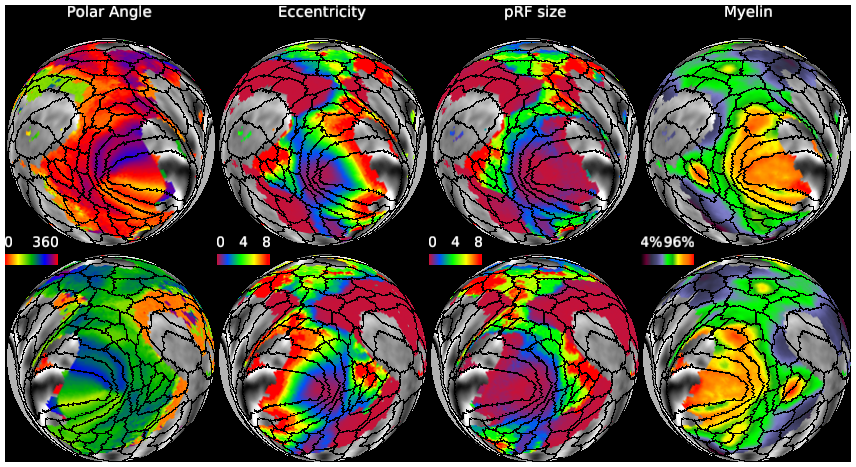
- Spheres: Angle; Eccentricity; pRF; Myelin (L-top, R-bottom)
-
Retinotopy_HCP_7T_Atlas.scene
SCENES: -
Retinotopy_HCP_7T_181_Fit2.scene
SCENES:- Spheres: Group avg Angle (0 - 360), Eccentricity, pRF, Myelin
- Flatmaps: Group Avg Angle, Eccentricity, pRF, Myelin
- Inflated: Group Avg Angle, Eccentricity, pRF, Myelin
- Midthickness: Group Avg Angle, Eccentricity, pRF, Myelin
- Subcortical retinotopy: LGN top row); Sup Colliculus (bottom row)
- Spheres: Individual-subject polar angle, eccentricity, pRF, myelin
- Spheres: Angle; Eccentricity; pRF; Myelin (L-top, R-bottom)
-
Retinotopy_HCP_7T_181_Fit3.scene
SCENES:- Spheres: Group avg Angle (0 - 360), Eccentricity, pRF, Myelin
- Flatmaps: Group Avg Angle, Eccentricity, pRF, Myelin
- Inflated: Group Avg Angle, Eccentricity, pRF, Myelin
- Midthickness: Group Avg Angle, Eccentricity, pRF, Myelin
- Subcortical retinotopy: LGN top row); Sup Colliculus (bottom row)
- Spheres: Individual-subject polar angle, eccentricity, pRF, myelin
- Spheres: Angle; Eccentricity; pRF; Myelin (L-top, R-bottom)
-
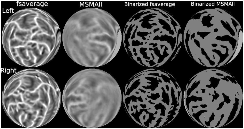
Retinotopy_HCP_7T_181_fsaverage.scene
SCENES: -
Retinotopy_HCP_7T_90_Fit1.scene
SCENES:- Spheres: Group avg Angle (0 - 360), Eccentricity, pRF, Myelin
- Flatmaps: Group Avg Angle, Eccentricity, pRF, Myelin
- Inflated: Group Avg Angle, Eccentricity, pRF, Myelin
- Midthickness: Group Avg Angle, Eccentricity, pRF, Myelin
- Subcortical retinotopy: LGN top row); Sup Colliculus (bottom row)
- Spheres: Angle; Eccentricity; pRF; Myelin (L-top, R-bottom)
-
Retinotopy_HCP_7T_91_Fit1.scene
SCENES:- Spheres: Group avg Angle (0 - 360), Eccentricity, pRF, Myelin
- Flatmaps: Group Avg Angle, Eccentricity, pRF, Myelin
- Inflated: Group Avg Angle, Eccentricity, pRF, Myelin
- Midthickness: Group Avg Angle, Eccentricity, pRF, Myelin
- Subcortical retinotopy: LGN top row); Sup Colliculus (bottom row)
- Spheres: Angle; Eccentricity; pRF; Myelin (L-top, R-bottom)
-
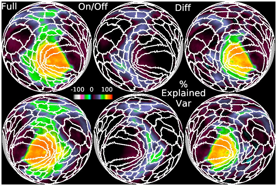
Retinotopy_HCP_7T_181_Fit1_R2.scene
SCENES: