FULL TITLE:
A Domain-general Cognitive Core defined in Multimodally Parcellated Human Cortex
SPECIES:
Human
ABSTRACT:
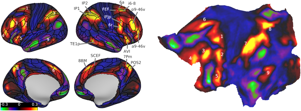
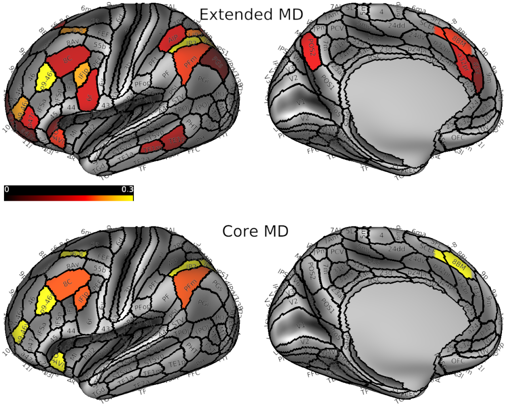
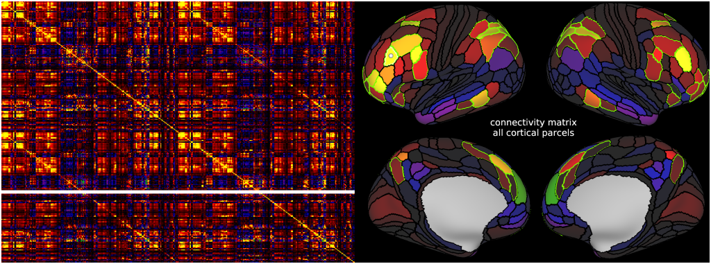
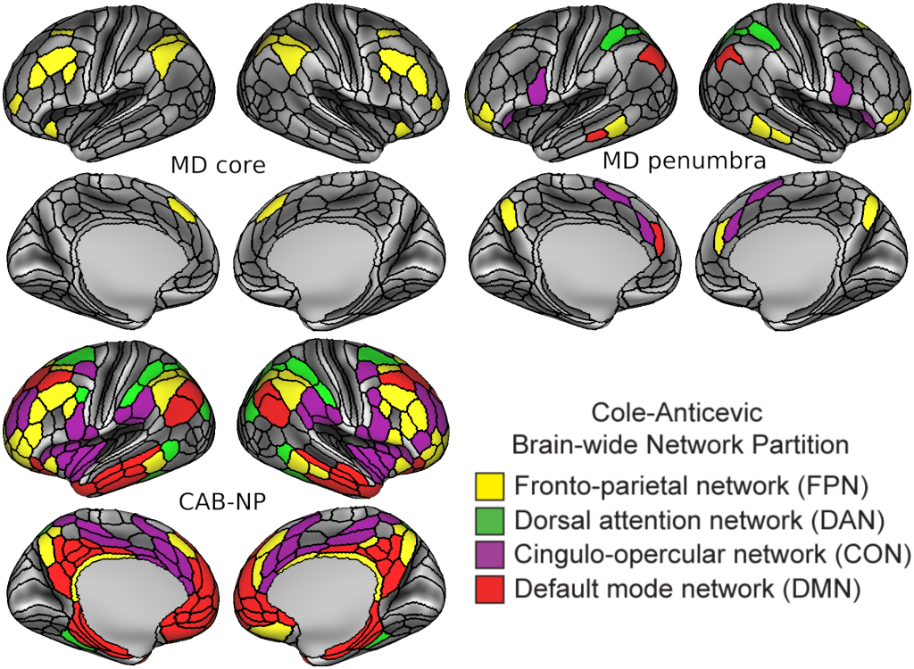
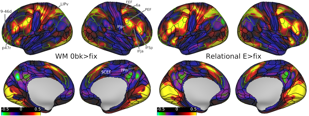
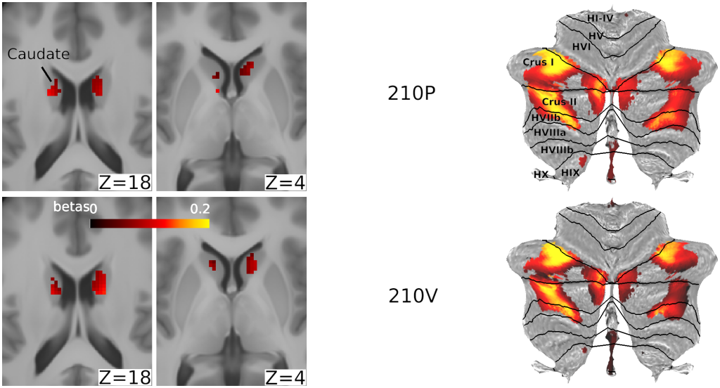
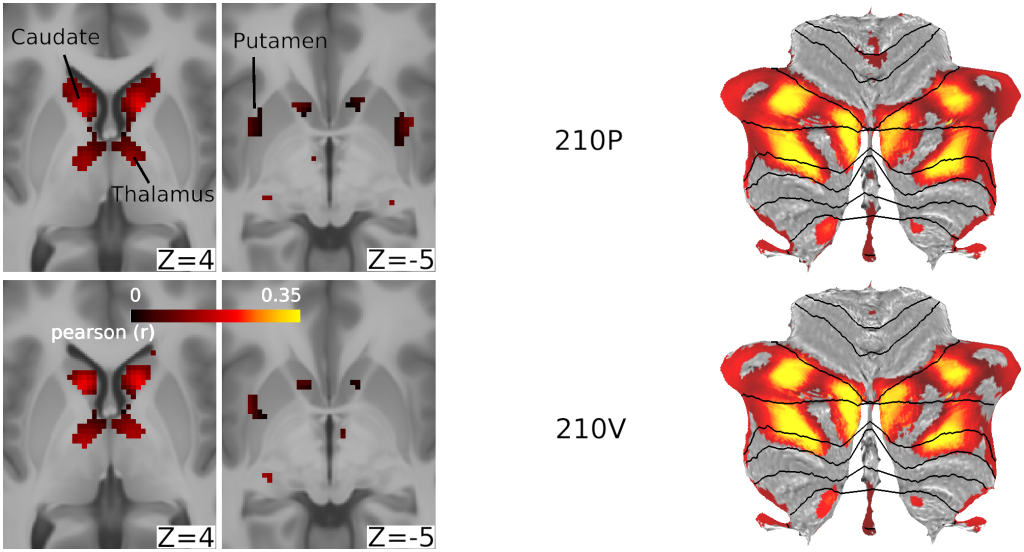
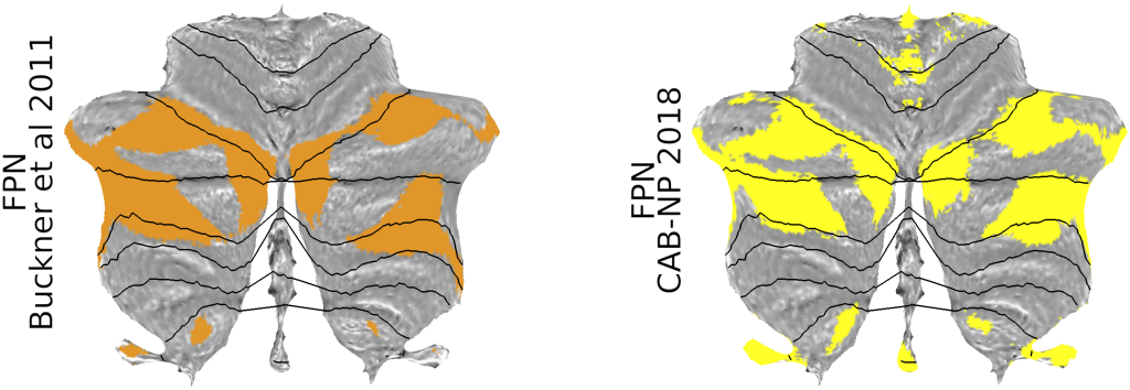
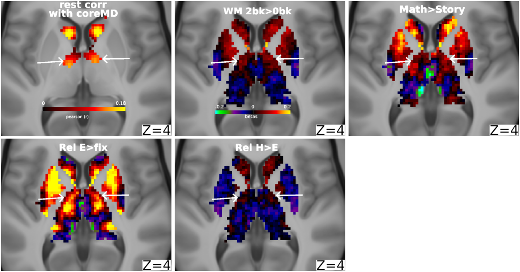
Numerous brain imaging studies identified a domain-general or “multiple-demand” (MD) activation pattern accompanying many tasks and may play a core role in cognitive control. Though this finding is well established, the limited spatial localization provided by traditional imaging methods precluded a consensus regarding the precise anatomy, functional differentiation and connectivity of the MD system. To address these limitations, we used data from 449 subjects from the Human Connectome Project, with cortex of each individual parcellated using neurobiologically grounded multi-modal MRI features. The conjunction of three cognitive contrasts reveals a core of 10 widely distributed MD parcels per hemisphere that are most strongly activated and functionally interconnected, surrounded by a penumbra of 17 additional areas. Outside cerebral cortex, MD activation is most prominent in the caudate and cerebellum. Comparison with canonical resting state networks shows MD regions concentrated in the fronto-parietal network but also engaging three other networks. MD activations show modest relative task preferences accompanying strong co-recruitment. With distributed anatomical organization, mosaic functional preferences, and strong interconnectivity, we suggest MD regions are well positioned to integrate and assemble the diverse components of cognitive operations. Our precise delineation of MD regions provides a basis for refined analyses of their functions.
PUBLICATION:
Cerebral Cortex
- DOI:
10.1093/cercor/bhaa023
- Moataz Assem
- Matthew F. Glasser
- David C. Van Essen
- John Duncan
- University of Oxford
- University of Cambridge
- St. Luke's Hospital
- Washington University in St. Louis
-
Assem_et_al 2020_DomainGeneralCognitiveCore.scene
SCENES:- Figure1_MDdense
- Figure2_MDparcels
- Figure3_MD_rest_connectivity
- Figure4_MD_in_CAB-NP
- Figure6e_MD_functional_preferences
- Figure7_MD_easydemand
- Figure8a_task_MD_subcortex_cerebellum
- Figure8b_rest_MD_subcortex_cerebellum
- Figure8c_FPN_cerebellum
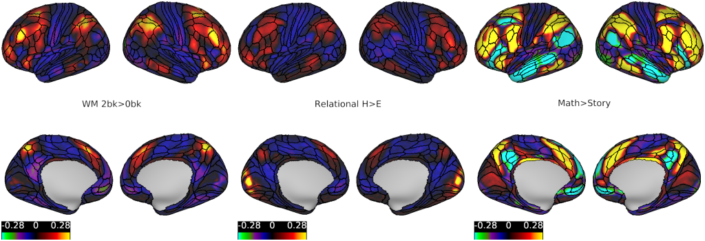
- FigureS1_separate_contrasts
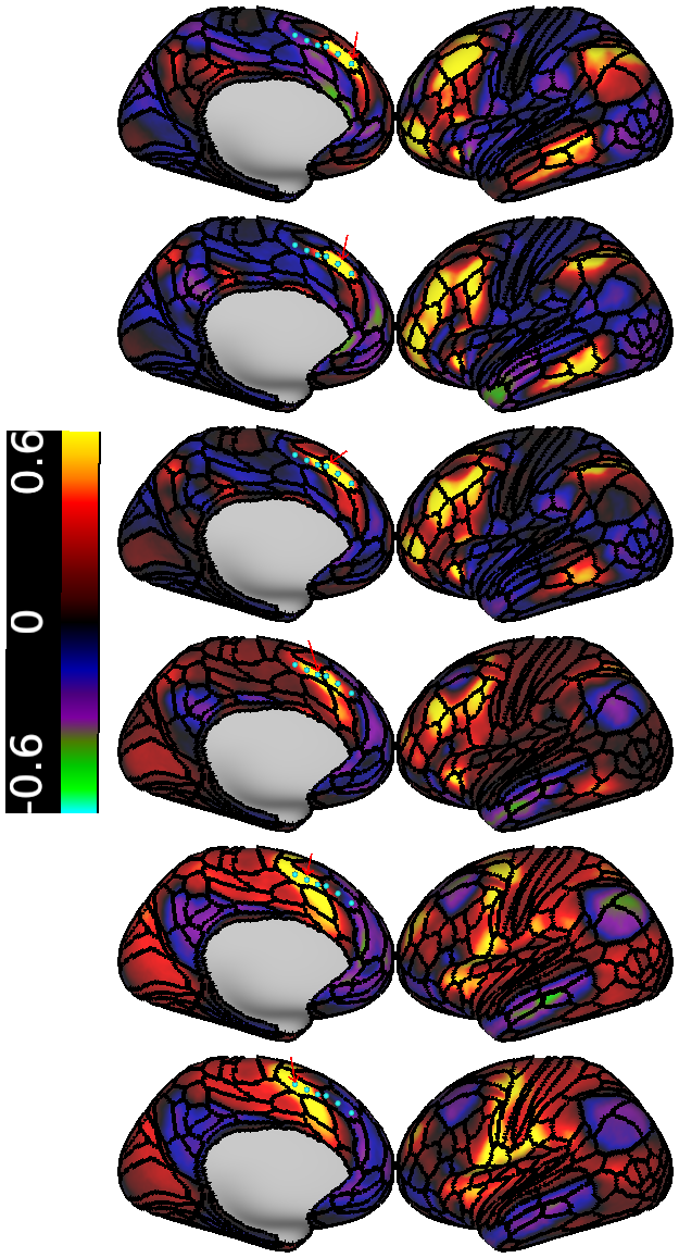
- FigureS2b_8BM_SCEF_seeds
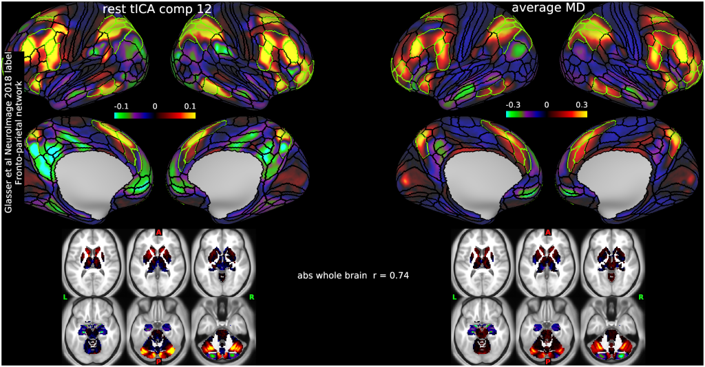
- FigureS3_top_MD_rest_temporalICA
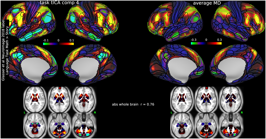
- FigureS3_bottom_MD_task_temporalICA
- FigureS5_MD_subcortical

