

FULL TITLE:
Multimodal 3D atlas of the macaque monkey inferior parietal lobe
SPECIES:
Macaque
DESCRIPTION:
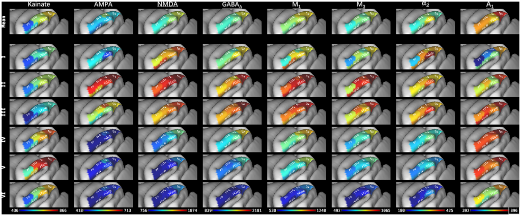
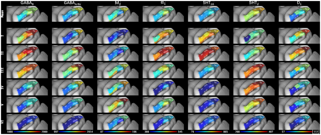
Parcellation scheme of the macaque inferior parietal lobule projected onto the lateral view of the Yerkes19 surface (Donahue et al., 2016). 6 cyto- and receptor architectonically distinc areas were ideintified in this brain region: four areas are located at different caudo-rostral levels on the IPL convexity (from caudal to rostral, areas Opt, PG, PFG, and PF); two additional areas in the parietal operculum (areas PGop caudally and PFop rostrally). The absolute mean areal densities and laminar densities of receptors for glutamate (AMPA, kainate, NMDA), GABA (GABAA, GABAB, GABAA associated benzodiazepine (GABAA/BZ) binding sites), acetylcholine (M1, M2, M3), norepinephrine (alpha1, alpha2), serotonin (5-HT1A, 5-HT2), dopamine (D1) and adenosine (A1) have been projected onto the corresponding area. Color bars code for receptor densities in fmol/mg protein.
ABSTRACT:
The macaque monkey inferior parietal lobe (IPL) is a structurally heterogeneous brain region, although the number of areas it contains and the anatomical/functional relationship of identified subdivisions remains controversial. Neurotransmitter receptor distribution patterns not only reveal the position of the cortical borders, but also segregate areas associated to different functional systems. Thus we carried out a multimodal quantitative analysis of the cyto- and receptor architecture of the macaque IPL to determine the number and extent of distinct areas it encompasses. We identified four areas on the IPL convexity arranged in a caudo-rostral sequence, as well as two areas in the parietal operculum, which we projected onto the Yerkes19 surface. We found rostral areas to have relatively smaller receptor fingerprints than the caudal ones, which is in an agreement with the functional gradient along the caudo-rostral axis described in previous studies. The hierarchical analysis segregated IPL areas into two clusters: the caudal one, contains areas involved in multisensory integration and visual-motor functions, and rostral cluster, encompasses areas active during motor planning and action-related functions. The results of the present study provide novel insights into clarifying the homologies between human and macaque IPL areas. The ensuing 3D map of the macaque IPL, and the receptor fingerprints are made publicly available to the neuroscientific community via the Human Brain Project and BALSA repositories for future cyto- and/or receptor architectonically driven analyses of functional imaging studies in non-human primates.
PUBLICATION:
NeuroImage
- DOI:
10.1016/j.neuroimage.2021.117843
- PMID:
33577936
- Meiqi Niu
- Lucija Rapan
- Thomas Funck
- Sean Froudist-Walsh
- Ling Zhao
- Karl Zilles
- Nicola Palomero-Gallagher
- Heinrich-Heine University
- New York University
- Medical Faculty, RWTH Aachen
- Research Centre Jülich