
Scene Preview
study:
Receptor architecture of macaque inferior parietal lobe
SCENE FILE:
Figure 6
SCENE:
Figure 6
DESCRIPTION:
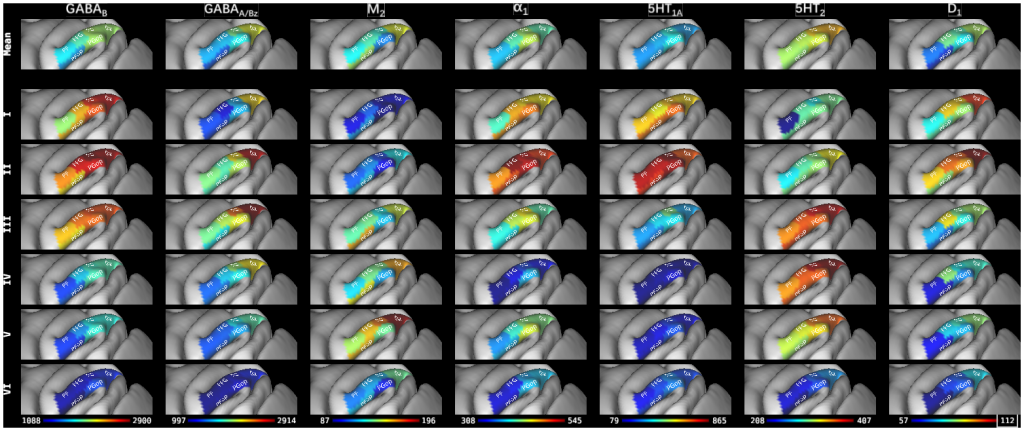
Figure 6. Parcellation scheme of the macaque inferior parietal lobule projected onto the lateral views of the Yerkes19 surface (Donahue et al., 2016). 6 cyto- and receptor architectonically distinct areas were identified in this brain region: four areas are located at different caudo-rostral levels on the IPL convexity (i.e. areas Opt, PG, PFG, and PF); two additional areas in the parietal operculum (i.e. areas PGop and PFop). The absolute mean areal densities and laminar densities of receptors for GABA (GABAB, GABAA associated benzodiazepine (GABAA/BZ) binding sites), acetylcholine (M2), norepinephrine (alpha1), serotonin (5-HT1A, 5-HT2), and adenosine (A1) have been projected onto the corresponding area. Color bars code for receptor densities in fmol/mg protein.
TAGS:
Atlas:Yerkes 19, Species:Macaque