
Scene Preview
study:
Receptor architecture of macaque inferior parietal lobe
SCENE FILE:
Figure S12
SCENE:
Figure S12
DESCRIPTION:
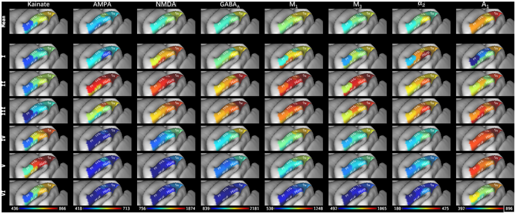
Figure S12. Parcellation scheme of the macaque inferior parietal lobule projected onto the lateral views of the Yerkes19 surface (Donahue et al., 2016). 6 cyto- and receptor architectonically distinct areas were identified in this brain region: four areas are located at different caudo-rostral levels on the IPL convexity (i.e. areas Opt, PG, PFG, and PF); two additional areas in the parietal operculum (i.e. areas PGop and PFop). The absolute mean areal densities and laminar densities of receptors for glutamate (AMPA, kainate, NMDA), GABA (GABAA), acetylcholine (M1, M3), norepinephrine (alpha2) and adenosine (A1) have been projected onto the corresponding area. Color bars code for receptor densities in fmol/mg protein.
TAGS:
Atlas:Yerkes 19, Species:Macaque