FULL TITLE:
The Detailed Organization of the Human Cerebellum Estimated by Intrinsic Functional Connectivity Within the Individual
SPECIES:
Human
DESCRIPTION:
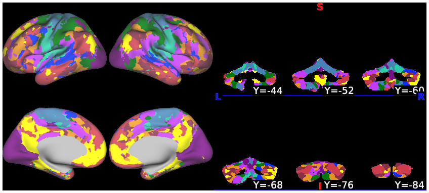
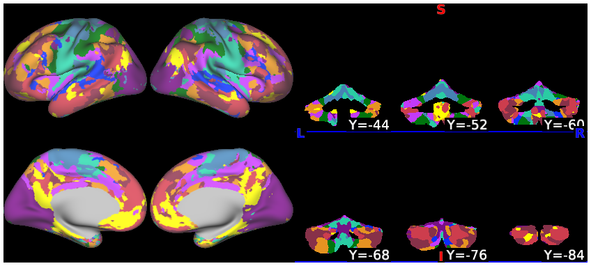
Parcellations for both individuals and seed maps in the paper are included in this study. Code is available at https://github.com/ThomasYeoLab/CBIG/tree/master/stable_projects/
brain_parcellation/Xue2021_IndCerebellum
ABSTRACT:
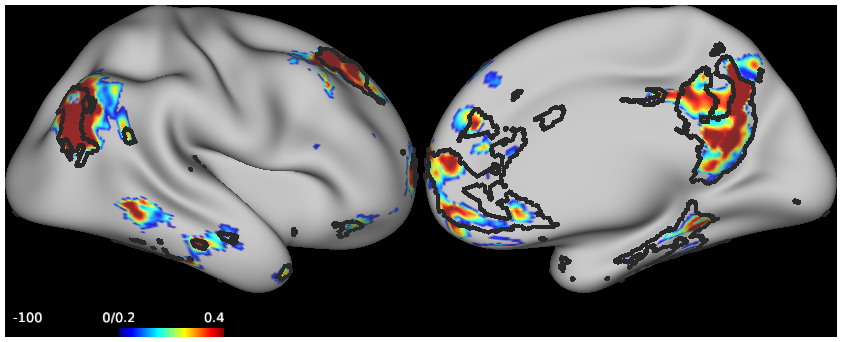
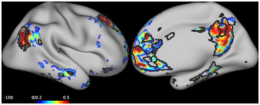
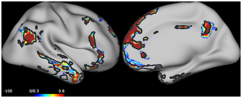
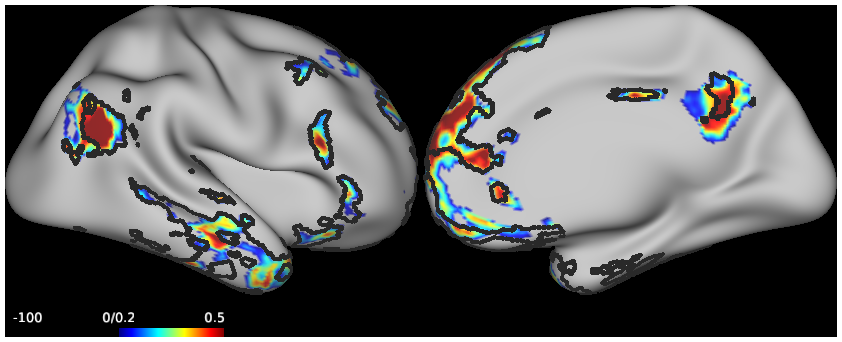
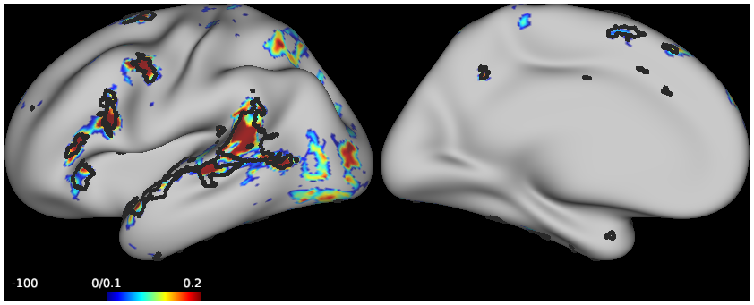
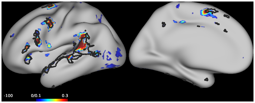
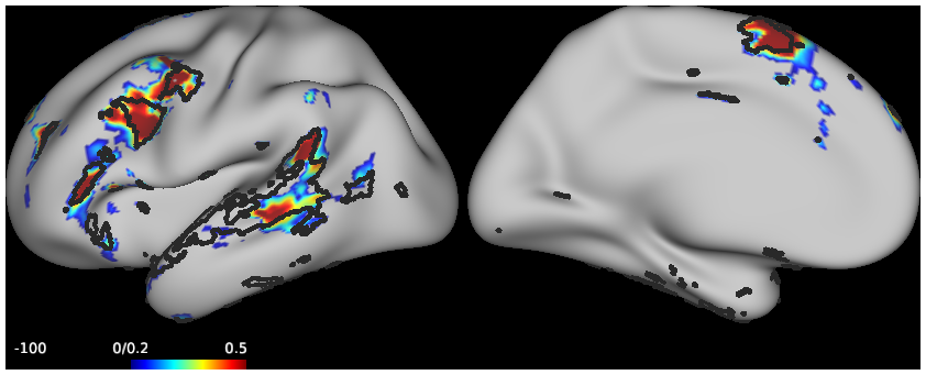
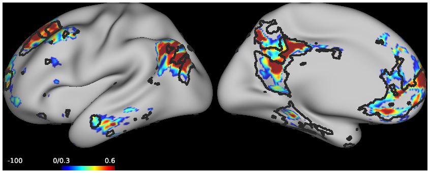
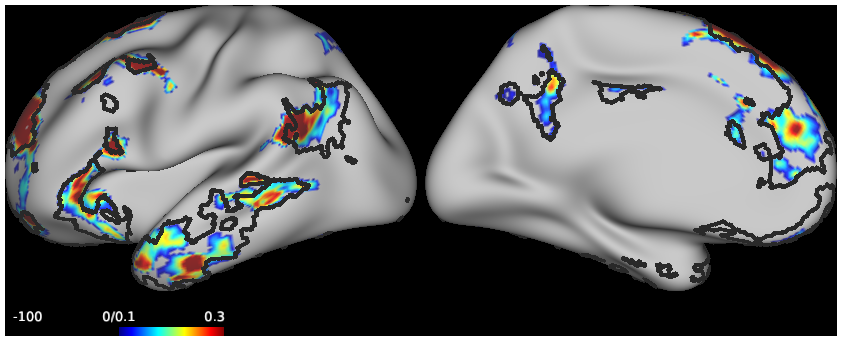
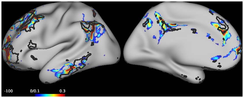
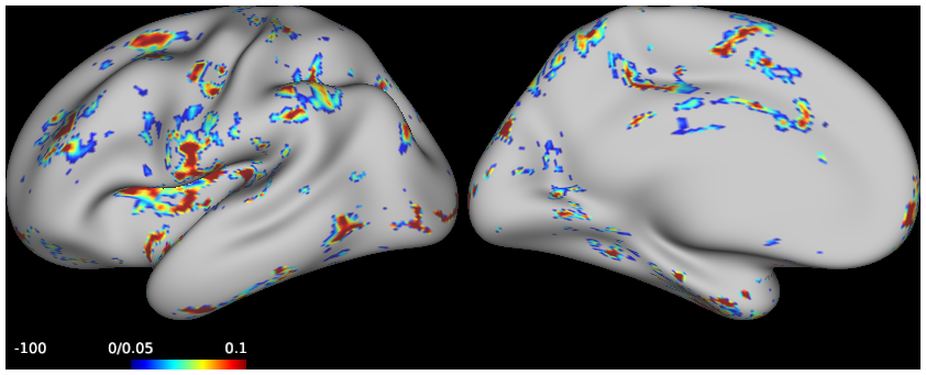
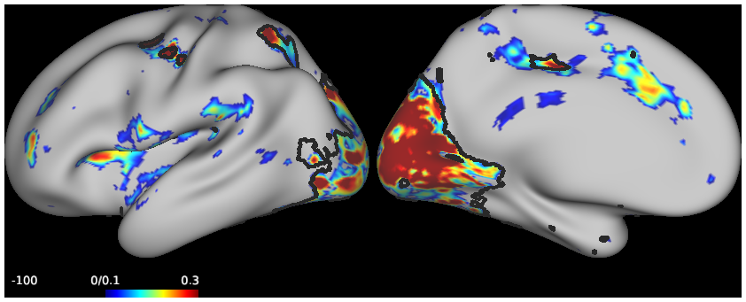
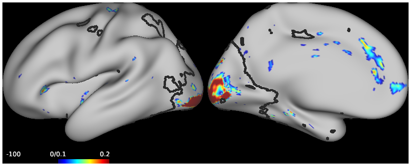
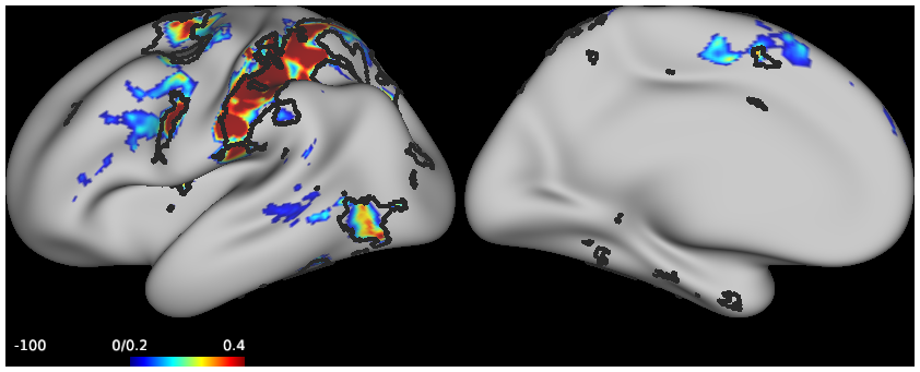
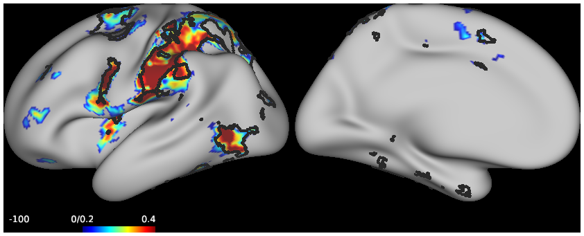
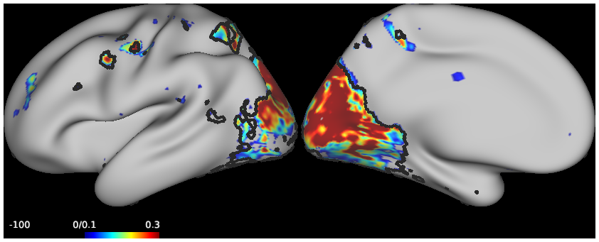
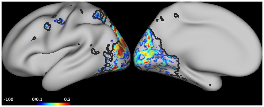
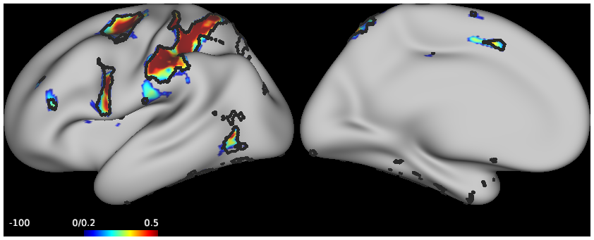
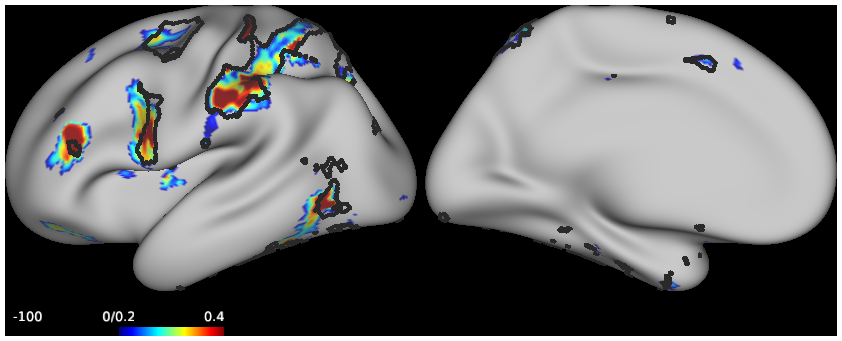
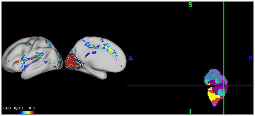
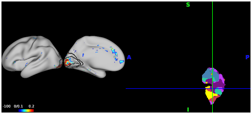
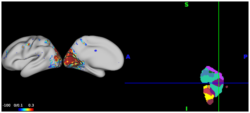
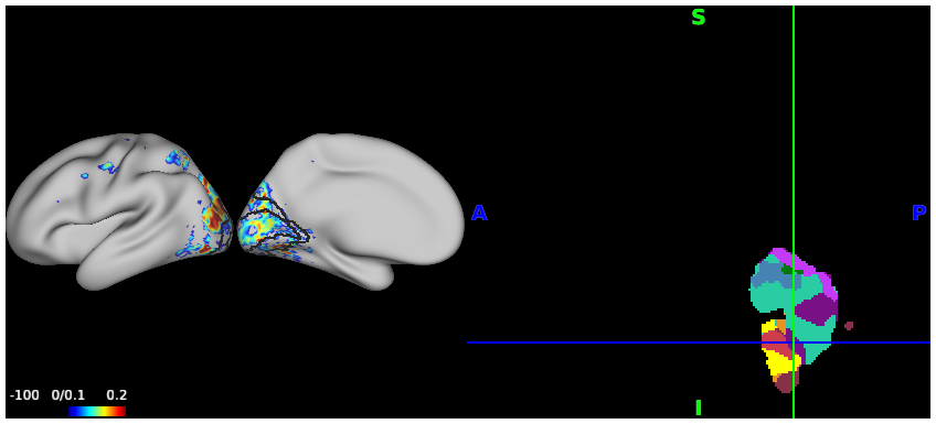
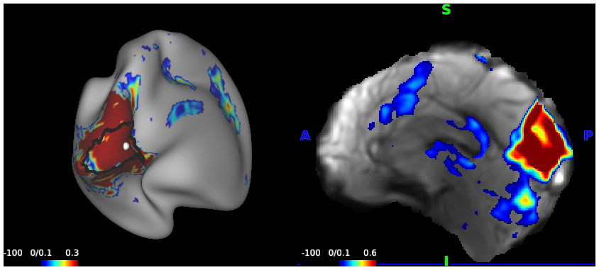
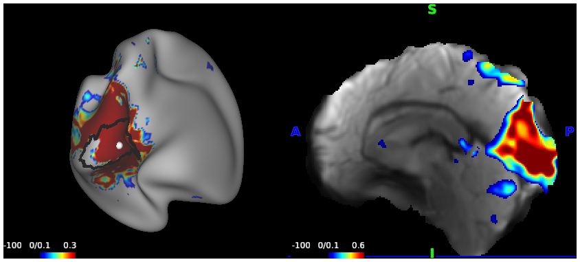
Distinct regions of the cerebellum connect to separate regions of the cerebral cortex forming a complex topography. While cerebellar organization has been examined in group-averaged data, study of individuals provides an opportunity to discover features that emerge at a higher spatial resolution. Here functional connectivity MRI was used to examine the cerebellum of two intensively-sampled individuals (each scanned 31 times). Connectivity to somatomotor cortex showed the expected crossed laterality and topography of the body maps. A surprising discovery was connectivity to the primary visual cortex along the vermis with evidence for representation of the central field. Within the hemispheres, each individual displayed a hierarchical progression from the inverted anterior lobe somatomotor map through to higher-order association zones. The hierarchy ended at Crus I/II and then progressed in reverse order through to the upright somatomotor map in the posterior lobe. Evidence for a third set of networks was found in the most posterior extent of the cerebellum. Detailed analysis of the higher-order association networks revealed robust representations of two distinct networks linked to the default network, multiple networks linked to cognitive control, as well as a separate representation of a language network. While idiosyncratic spatial details emerged between subjects, each network could be detected in both individuals, and seed regions placed within the cerebellum recapitulated the full extent of the spatially-specific cerebral networks. The observation of multiple networks in juxtaposed regions at the Crus I/II apex confirms the importance of this zone to higher-order cognitive function and reveals new organizational details.
PUBLICATION:
Journal of Neurophysiology
- DOI:
10.1152/jn.00561.2020
- Aihuiping Xue
- Ru Kong
- Qing Yang
- Mark C. Eldaief
- Peter Angeli
- Lauren M. DiNicola
- Rodrigo M. Braga
- Randy L. Buckner
- B.T. Thomas Yeo
- Massachusetts General Hospital
- NUS Graduate School for Integrative Sciences and Engineering, National University of Singapore, Singapore, 117574, Singapore
- Department of Neurology, Northwestern University Feinberg School of Medicine, Chicago, IL 60611, USA
- Harvard University
-
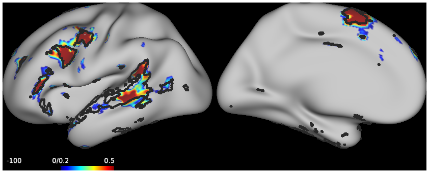
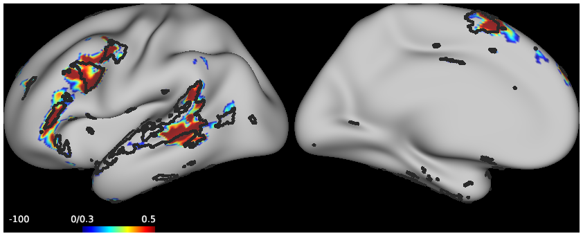
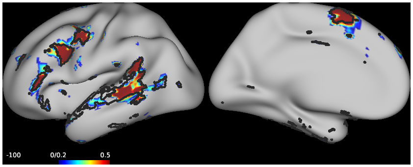
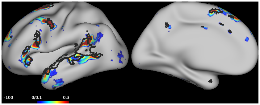
Figures.scene
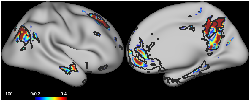
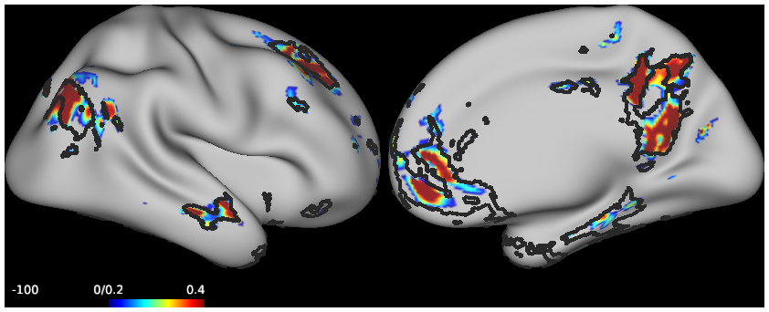
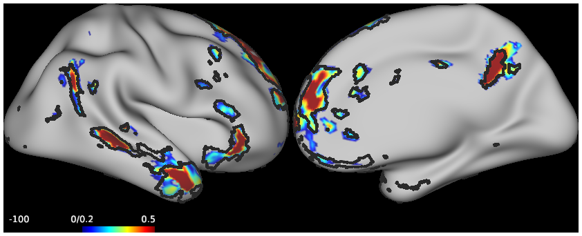
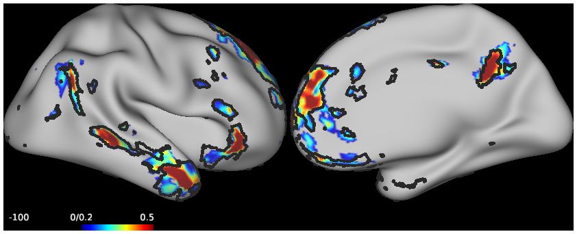
SCENES:- Figure12_Sub1_DNA_1
- Figure12_Sub1_DNA_2
- Figure12_Sub1_DNB_1
- Figure12_Sub1_DNB_2
- Figure_12_Sub2_DNA_1
- Figure12_Sub2_DNA_2
- Figure12_Sub2_DNB_1
- Figure12_Sub2_DNB_2
- Figure13_Sub1_Language_1
- Figure13_Sub1_Language_2
- Figure13_Sub1_Language_3
- Figure13_Sub2_Language1
- Figure13_Sub2_Language_2
- Figure13_Sub2_Language_3
- Figure14_Sub1_Language
- Figure14_Sub1_DNA
- Figure14_Sub2_DNB
- Figure14_Sub2_ControlB
- Figure15
- Figure16_Sub1_Visual_1
- Figure16_Sub1_Visual_2
- Figure16_Sub1_Dorsal_1
- Figure16_Sub1_Dorsal_2
- Figure16_Sub2_Visual_1
- Figure16_Sub2_Visual_2
- Figure16_Sub2_Dorsal_1
- Figure16_Sub2_Dorsal_2
- Figure18_Sub1_Peripheral
- Figure18_Sub1_Central
- Figure18_Sub2_Peripheral
- Figure18_Sub2_Central
- Figure19_Sub1
- Figure19_Sub2
-
Parcellation.scene
SCENES:

