FULL TITLE:
Cortical adaptation of the night monkey to a nocturnal niche environment: A comparative non-invasive T1w/T2w myelin study
SPECIES:
Marmoset, Macaque
ABSTRACT:
Night monkeys (Aotus) are the only genus of monkeys within the Simian lineage that successfully occupy the nocturnal niche environment. Their behavior is supported by their sensory organs’ unique morphological features; however, little is known about their evolutionary adaptations in sensory regions of the cerebral cortex. Here, we investigate this question by exploring the cortical organization of night monkeys using high-resolution in-vivo brain MRI and comparative cortical-surface T1w/T2w myeloarchitectonic mapping. Our results show that the night monkey cerebral cortex has a similar organization of cortical myelin distribution compared to the diurnal macaque and marmoset monkeys. T1w/T2w myelin and its gradient allowed us to parcellate high myelin areas, middle temporal complex (MT+MST) and auditory cortex, and low myelin area, Brodmann area 7 (BA7) in three species, despite the difference in gyrifications. Relative to the total cortical surface area, those of MT+MST and the auditory cortex are significantly larger in night monkeys than diurnal monkeys. In contrast, the relative area of BA7 is expanded in proportion to brain size across species. We propose that the selective expansion of sensory areas dedicated to visual motion and auditory processing in night monkeys may reflect cortical adaptations to the nocturnal niche environment.
PUBLICATION:
Brain Function and Structure
- Takuro Ikeda
- Joonas A. Autio
- Akihiro Kawasaki
- Chiho Takeda
- Takayuki Ose
- Masahiko Takada
- David C. Van Essen
- Matthew F. Glasser
- Takuya Hayashi
- Washington University in St. Louis
- RIKEN
- Kyoto University
-
MainFigures.scene
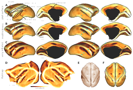
SCENES:- Figure 1. Surfaces models of night monkey cerebral cortex
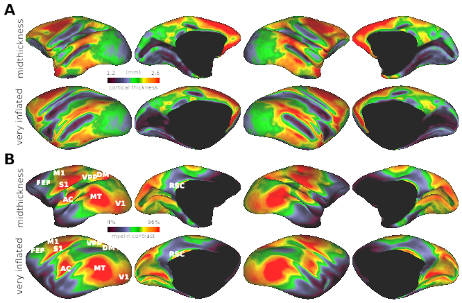
- Figure 2. Thickness and myeloarchitecture in the night monkey cerebral cortex. (Panel 1)
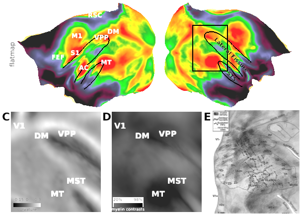
- Figure 2. Thickness and myeloarchitecture in the night monkey cerebral cortex. (Panel 2)
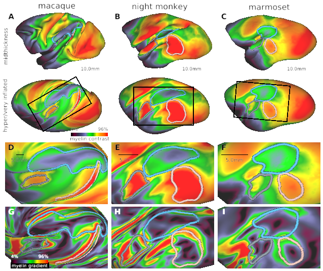
- Figure 3. Interspecies comparison of myeloarchitecture in parieto-temporal cortex.


