
Scene Preview
study:
Non-invasive myelin mapping of the night monkey
SCENE FILE:
MainFigures
SCENE:
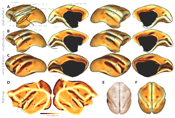
Figure 1. Surfaces models of night monkey cerebral cortex
DESCRIPTION:
Cortical curvature displayed on (A) pial, (B) midthickness and (C) very inflated surfaces, and (D) a flatmap. Three sulci (lateral fissure, superior temporal and cingulate sulcus) and four dimples (principal, arcuate and central dimple) were consistently identified in all of the animals (N=9). Dorsal views of (E) postmortem brain (modified image from http://neurosciencelibrary.org/) and (F) reconstructed pial surface. Red dots are placed by a regular interval on the 'anatomical coordinates' of midthickness surface. Note that the corresponding red dots are located in a distorted manner in the very-inflated and flat surfaces. The cyan dots in the right hemisphere are vertices with the same ID contralateral to the red dots in the left hemisphere demonstrate symmetrical reconstruction of the cortical surfaces.
TAGS: