
Scene Preview
study:
Non-invasive myelin mapping of the night monkey
SCENE FILE:
MainFigures
SCENE:
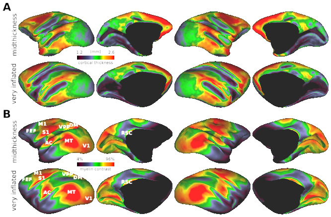
Figure 2. Thickness and myeloarchitecture in the night monkey cerebral cortex.
(Panel 1)
DESCRIPTION:
(A) Cortical thickness distribution displayed on midthickness (upper) and very inflated surfaces (lower panel). (B) T1w/T2w myelin contrast displayed on midthickness (upper panel), very inflated surface (lower panel), and flatmap (lower). The zoomed view of (C) curvature and (D) T1w/T2w myelin contrast in the parieto-temporal cortical area (the black rectangle in flatmap) in comparison to (E) histological flat-mounted section of myelin stain (Sereno et al. 2015). The image intensity indicates myelin density (bright and dark indicate low and high density, respectively). Note the spatial similarity between T1w/T2w myelin contrast and the histological myelin density. Abbreviations: AC: auditory cortex; FEF: frontal eye field; DM: dorsomedial visual area; MT: Middle temporal area; RSC: retrosplenial cortex; S1: primary somatosensory cortex; STS: superior temporal sulcus; V1: primary visual cortex; VPP: ventroposterior parietal area.
TAGS: