
Scene Preview
Scene: Figure 3A: Human PFC Border Variability
study:
PFC in Humans Relative to Nonhuman Primates
SCENE FILE:
DonahueEtAl2018_PNAS
SCENE:
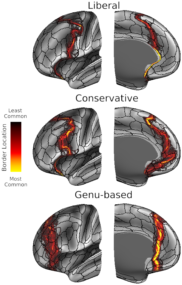
Figure 3A: Human PFC Border Variability
DESCRIPTION:
PFC border probability maps displayed on inflated left hemisphere atlas surfaces of the frontal lobe (lateral aspect on left and medial aspect on right of each pair), overlaid on group-average sulcal depth maps. Human liberal and conservative PFC borders were created using individual subject parcellations, resulting in pronounced intersubject variance on the group-average surface.
TAGS:
Surface Mesh:164k fs LR, Registration:MSMAll, Species:Human