
Scene Preview
study:
Comparative Connectomics of the Primate Social Brain
SCENE FILE:
Macaque_Human_Homologs
SCENE:
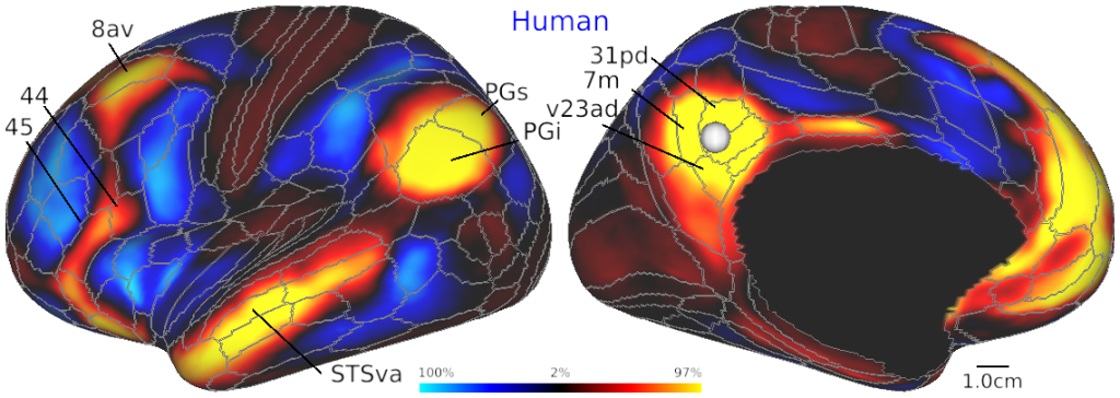
Human pC/PCC seed FC (ID=11635)
DESCRIPTION:
Putative homologs of functional networks in the left hemispheres of macaque (top) and human (bottom) cerebral cortices. (A) Dense functional connectivity (FC) seeded from the left ventral premotor (vPM) cortex (areas F5a and 6r in macaque and human, respectively) reveals a fronto-parietal network mostly localized near to the primary sensory cortex. Note that positive FC extends over premotor, inferior parietal and insular cortices whereas anticorrelation extends over temporal and prefrontal cortices. (B) FC seeded from the left posterior cingulate cortex/precuneus (pC/PCC) reveals another fronto-parieto-temporal association network that exhibits positive FC in the angular gyrus (parietal area G inferior, PGi), parietal area G superior (PGs) and medial prefrontal cortices. Interestingly, the vPM and pC/PCC FC are spatially anticorrelated in both species. Annotations of cortical areas and boundaries (grey color) are according to M132 (Markov et al., 2014) and the Young Adult Human Connectome Project (YA-HCP) (Glasser et al., 2016b) parcellations in macaque and human, respectively. Human data is from the YA-HCP S1200 Release whereas the macaque data (N=30) was obtained using standardized HCP-style data acquisition and preprocessing (Autio et al., 2020a; Glasser et al., 2013; Hayashi et al., 2021, 2019) and standardized in a CIFTI greyordinate space in each species (see Section 4.3). Fig. 2b was reproduced from Hayashi et al., 2021.Note that while the human HCP_MMP cortical parcellation was created based on multimodal MRI data including functional connectome, the macaque parcellation is created based on neuroanatomy but often disagrees with functional maps in some of the areas, e.g. posterior cingulate cortex (Vogt et al. 1987, 2006).
TAGS:
Species:Human