
Scene Preview
study:
A Multi-modal Parcellation of Human Cerebral Cortex
SCENE FILE:
Glasser_et_al_2016_HCP_MMP1.0_1_MainText
SCENE:
Figure 1
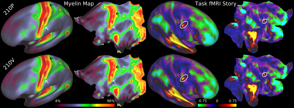
DESCRIPTION:
Consistency of fine spatial details in independent group averages. Relative myelin content maps (left hemisphere) and task fMRI contrast beta maps from the LANGUAGE story contrast (right hemisphere) on inflated (columns 1 and 3) and flattened surfaces (columns 2 and 4). Rows 1 and 2 are the group averages of the 210P and 210V data sets, respectively. White and black arrows indicate consistent variations in myelin content within primary somatosensory cortex that are correlated with somatotopy (see Supplementary Neuroanatomical Results 6 and Supplementary Neuroanatomical Results Fig. 8). The white oval indicates a small, sharp, and reproducible feature in the right hemisphere of the LANGUAGE story contrast. Relative myelin content will hereafter be referred to as myelin (see legend of Supplementary Fig. 1 in Supplementary Results and Discussion 1.1).
TAGS:
Surface Mesh:32k fs LR, Registration:MSMAll, Species:Human, Modality:Myelin Map, Modality:T1-weighted, Modality:T2-weighted