
Scene Preview
study:
A Multi-modal Parcellation of Human Cerebral Cortex
SCENE FILE:
Glasser_et_al_2016_HCP_MMP1.0_4_SupplementaryNeuroanatomicalResults
SCENE:
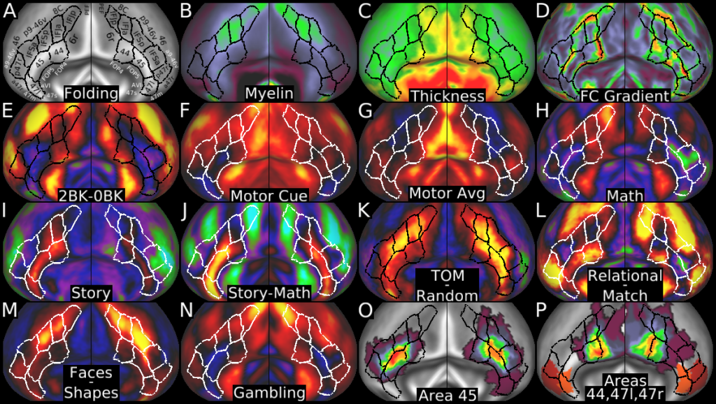
Figure 24
DESCRIPTION:
Figure 24 shows multi-modal information used to parcellate the inferior frontal cortex. Panel A shows the areas on a folding map. Panels B and C show myelin and cortical thickness maps. Panel D shows the resting state functional connectivity gradients. Panels E, F, G, and H show the working memory 2BK-0BK contrast, the MOTOR CUE contrast, the MOTOR AVG contrast, and the LANGUAGE MATH contrast. Panels I, J, K and L show the LANGUAGE STORY contrast, the STORY-MATH contrast, the SOCIAL TOM-RANDOM contrast, and the RELATIONAL-MATCH contrast. Panels M and N show the EMOTION FACES-SHAPES contrast and a GAMBLING primary contrast. Panel O shows the surface-based probabilistic map of area 45 from (Fischl et al., 2008). Panel P shows the surface-based probabilistic map of area 44 from (Fischl et al., 2008) and maps of area 47l and 47r from (Ongur et al., 2003). While the parcellation is reasonably symmetric (parcel boundaries in the same locations relative to folding and areal features) the probabilistic maps have some asymmetries, suggesting that registration drift may contribute to some of the mismatches.
TAGS:
Surface Mesh:32k fs LR, Registration:MSMAll, Species:Human, Modality:T1-weighted, Modality:T2-weighted, Modality:Myelin Map, Atlas:Conte69