
Scene Preview
study:
A Multi-modal Parcellation of Human Cerebral Cortex
SCENE FILE:
Glasser_et_al_2016_HCP_MMP1.0_2_SupplementaryResultsAndDiscussion
SCENE:
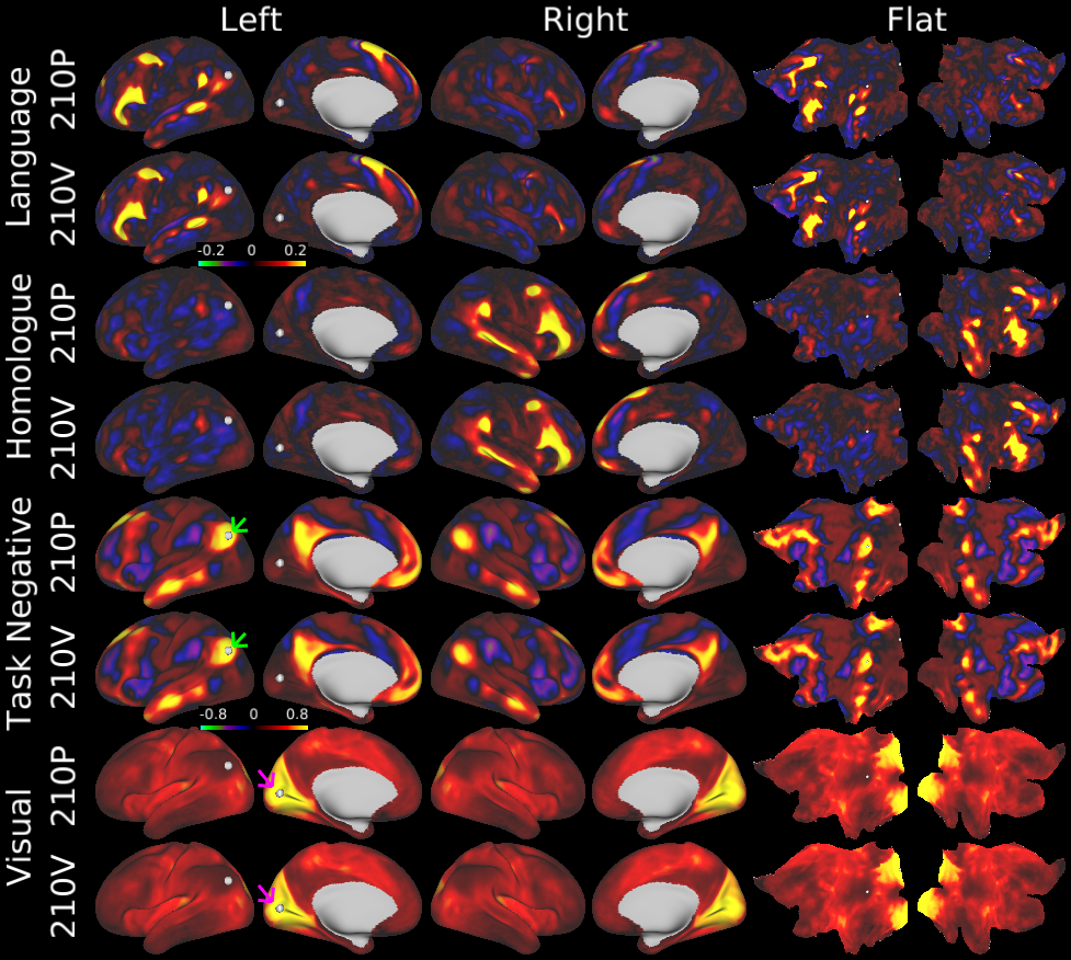
Figure 4
DESCRIPTION:
HCP group average resting-state fMRI reproducibility. Here we show the reproducibility of Resting State Networks (RSNs, ICA d=137) and dense Functional Connectivity maps. The odd rows are the 210P dataset and the even rows are the 210V dataset. The RSNs were produced using ICA on the 210P dataset and then weighted regression of the group spatial maps onto the individual subject dense timeseries to produce individual subject spatial maps in all 449 subjects. The individual subject maps were then separately averaged across the 210P and 210V groups. The top two rows show the left lateralized language network and the next two rows show its right hemisphere homologue, both scaled between beta=+/-0.2. The next two rows show dense functional connectivity (FC) of the bilateral task negative (default mode) network seeded from a posterior inferior parietal grayordinate (green arrow, white sphere on left lateral surface), which has strong anti-correlation with the task positive network. The last two rows show the early visual network seeded from a grayordinate in the center of the calcarine sulcus (purple arrow, white sphere on left medial surface), which shows positive correlation with the entire brain. The dense FC maps are scaled from r=-0.8 to 0.8.
TAGS:
Surface Mesh:32k fs LR, Registration:MSMAll, Species:Human, Modality:Myelin Map, Modality:T1-weighted, Modality:T2-weighted