
Scene Preview
study:
A Multi-modal Parcellation of Human Cerebral Cortex
SCENE FILE:
Glasser_et_al_2016_HCP_MMP1.0_4_SupplementaryNeuroanatomicalResults
SCENE:
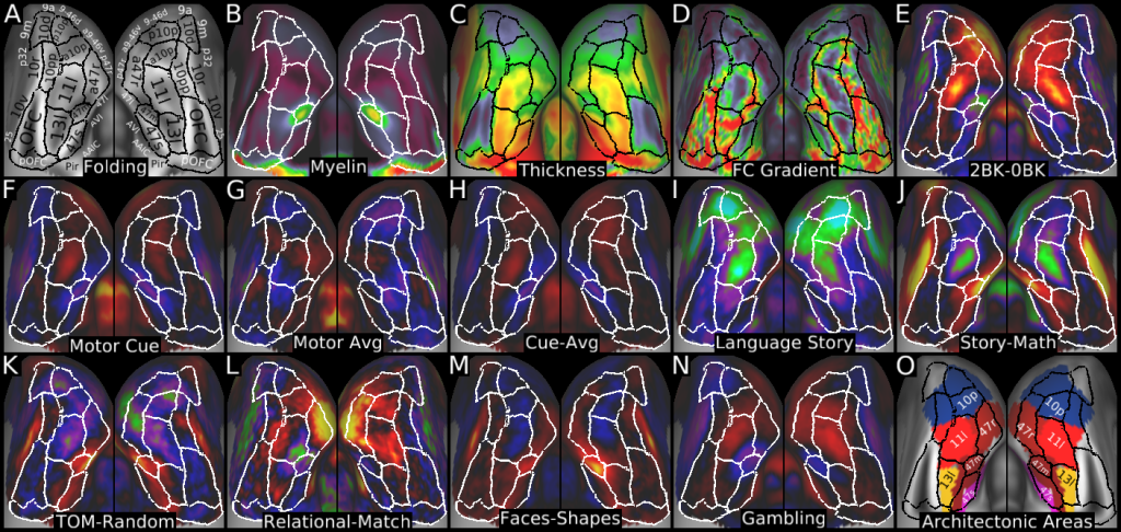
Figure 23
DESCRIPTION:
Figure 23 shows the multi-modal information that was used to parcellate the orbital and polar frontal cortex. Panel A shows the areas on a folding map. Panels B and C show myelin and cortical thickness maps. Panel D shows the resting state functional connectivity gradient. Panel E shows the working memory 2BK-0BK contrast. Panels F, G, and H show the MOTOR CUE, AVG, and CUE-AVG contrasts. Panels I and J show the LANGUAGE STORY and STORY-MATH contrasts. Panels K, L, M, and N show the TOM-RANDOM, RELATIONAL-MATCH, FACES-SHAPES, and GAMBLING primary contrasts. Panel O shows some areas (13l, 11l, 47m, 47s, 47r, and 10p) from the (Ongur et al., 2003) parcellation that were used to name areas in our parcellation.
TAGS:
Surface Mesh:32k fs LR, Registration:MSMAll, Species:Human, Modality:T1-weighted, Modality:T2-weighted, Modality:Myelin Map, Atlas:Conte69