
Scene Preview
study:
A Multi-modal Parcellation of Human Cerebral Cortex
SCENE FILE:
Glasser_et_al_2016_HCP_MMP1.0_4_SupplementaryNeuroanatomicalResults
SCENE:
Figure 9
DESCRIPTION:
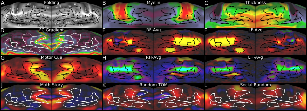
Figure 9 shows some of the multi-modal information used to delineate cortical areas in the paracentral lobular and mid cingulate cortical region. Panel A shows the areas delineated in this section overlaid on a folding map. Panels B and C show myelin and thickness maps. Panel D shows the resting state functional connectivity gradient. Panels E and F show the MOTOR RF-AVG and LF-AVG task fMRI contrasts (referred to as ?F-AVG in the text). Panel G shows the MOTOR CUE task contrast. Panels H and I show the RH-AVG and LH-AVG task contrasts (referred to as ?H-AVG in the text). Panels J, K and L show the MATH-STORY, RANDOM-TOM, and SOCIAL RANDOM task contrasts.
TAGS:
Modality:Myelin Map, Surface Mesh:32k fs LR, Registration:MSMAll, Species:Human, Modality:T1-weighted, Modality:T2-weighted