
Scene Preview
study:
Neurite imaging reveals microstructural variations in human cerebral cortical gray matter.
SCENE FILE:
Figures_NODDI
SCENE:
Fig1
DESCRIPTION:
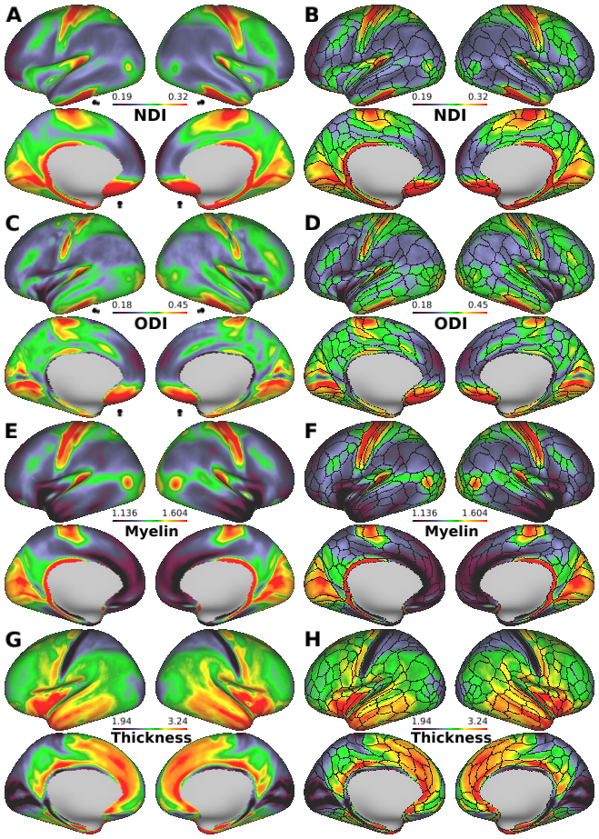
Diffusion MRI derived neurite properties, myelin, and cortical thickness maps of 505-subject average cerebral cortex. (A) Neurite density index (NDI), (C) orientation dispersion index (ODI), (E) myelin and (G) cortical thickness. (B, D, F and H) The imaging modalities superimpose with the boundaries of the Human Connectome Project (HCP) multi-modal parcellations, 210P MPM (black lines) (Glasser et al, 2016). NDI is relatively uniform within most of the parcels, and transitions in neurite properties often occur near parcel boundaries, whereas ODI is heterogenous in some parcellations, such as motor, somatosensory, and primary visual (see also Fig. 3). The black arrows (anterior and middle cranial fossa) indicate artifacts regions where the NDI and ODI values are overestimated because of low signal-to-noise ratio.
TAGS:
Surface Mesh:32k fs LR, Modality:Myelin Map, Registration:MSMAll, Species:Human