
Scene Preview
study:
Perspective for NHP_NNP
SCENE FILE:
Figures
SCENE:
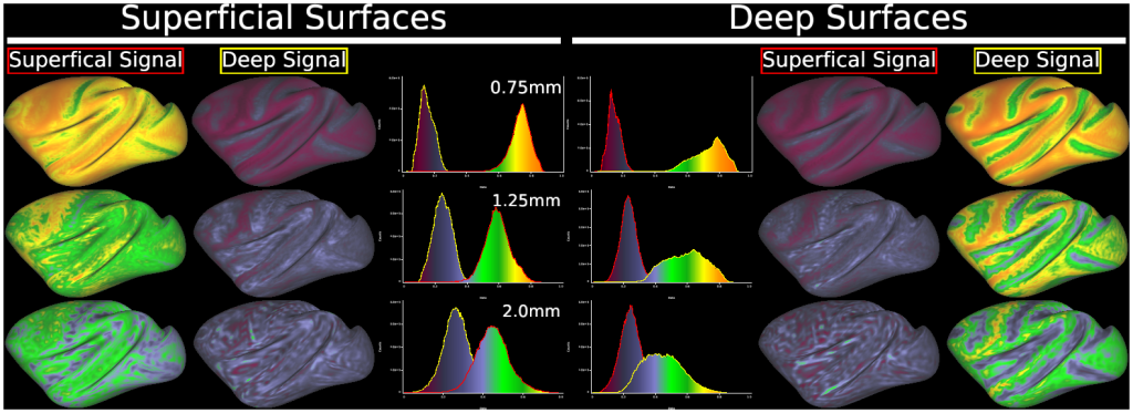
Fig. 8 Results of simulation for partial volume/geometric effects in ‘layer’ fMRI analysis at various resolutions in macaque cerebral cortex.
DESCRIPTION:
Simulation performed estimation of ‘layer 4’ surface for each subject of macaques (N=30) from Mac30BS, created fMRI-resolution volumes of simulated superficial and deep cortical signal (above and below layer 4, respectively), and re-mapped each layer’s signal onto the subject’s surfaces with ribbon mapping (pial to layer4, and layer4 to white), and these values were then averaged across subjects. In the corresponding layer/surface, a value closer to one indicates better response to layer-specific signal, where the values for the other layer indicate ‘spill-in’ of signals to the undesired layer. The layer 4 surface is approximated by the ‘equal volume method’ (locally, the same amount of cortical volume is superficial to the new surface as is deeper than it) (Van Essen and Maunsell, 1980). The use of conventional resolution in NHP (=2.0mm) results in significant loss or spill-out of signals in the corresponding layer, and spill-in into the other layer resulting in the overlap of histograms. NHP_NNP 3T protocol (1.25mm) was partly overlapped in histogram, and ultra-high field MRI protocol (0.75mm) showed distinct separation between peaks in the histograms, suggesting differentiation of signal between laminae in surface-based group analysis if one only considers geometric constraints (note that the separability of lamina also depends on the point-spread-function of BOLD fMRI or other fMRI approaches). See also Fig. S8 in (Coalson et al., 2018).
TAGS:
Surface Mesh:32k fs LR, Modality:T1-weighted, Atlas:Yerkes 19, Species:Macaque, Modality:Myelin Map, Modality:T2-weighted