
FULL TITLE:
Charting cortical layer-specific area boundaries using Gibbs’ ringing attenuated T1w/T2w-FLAIR myelin MRI
SPECIES:
Macaque
DESCRIPTION:
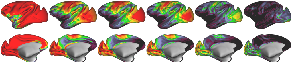
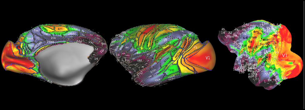
The equivolumetric layer scheme of macaque cerebral cortex used to delineate putative cortical area boundaries.
ABSTRACT:
Cortical areas have traditionally been defined by their distinctive layer cyto- and/or myelo- architecture using postmortem histology. Recent studies have delineated many areas by measuring overall cortical myelin content and its spatial gradients using T1w/T2w ratio MRI in living primates, including humans. While T1w/T2w studies of areal transitions might benefit from using the layer profile of this myelin-related contrast, a significant confound is Gibbs’ ringing artefact, which produces signal fluctuations resembling cortical layers. Here, we address these issues with a novel approach using cortical layer thickness-adjusted T1w/T2w-FLAIR imaging in the macaque cortex, which attenuates the amplification of opposite-phase Gibbs ringing seen in the conventional T1w/T2w ratio by exploiting the in-phase Gibbs pattern of T2w-FLAIR relative to T1w, thereby reducing ringing artefacts and improving intracortical myelin contrast. Whole-brain MRI measures were mapped onto twelve equivolumetric layers, and comparison with histological Gallyas myelin stains explains over 80% of the variance in the laminar T1w/T2w-FLAIR profiles, substantiating the validity of the method. Layer-specific sharp myeloarchitectonic transitions were then identified using spatial gradients, resulting in a putative 182 area/subarea partition of the macaque cerebral cortex. The myelin maps exhibit notably high homology with those in humans, suggesting cortical myelin shares a similar developmental program across species. Altogether, our approach provides a non-invasive means for precision mapping of laminar myeloarchitecture in the primate cerebral cortex, advancing the pioneering work of classical neuroanatomists.
PUBLICATION:
Imaging Neuroscience
-
Autio2026_wwmGp.scene
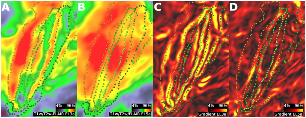
SCENES:- Figure 2A. Validating Laminar Myelin Mapping.
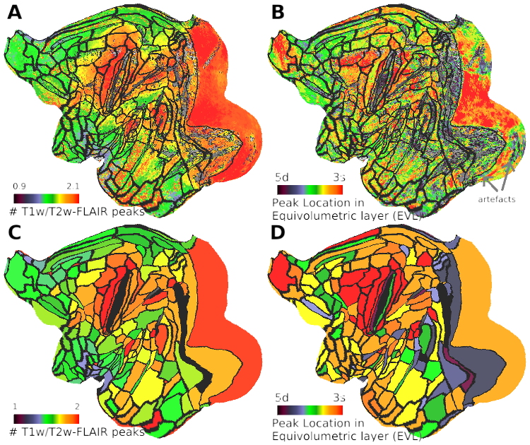
- Figure3. Charting cortical area boundaries using cortical layer-specific myeloarchitectonic transitions.
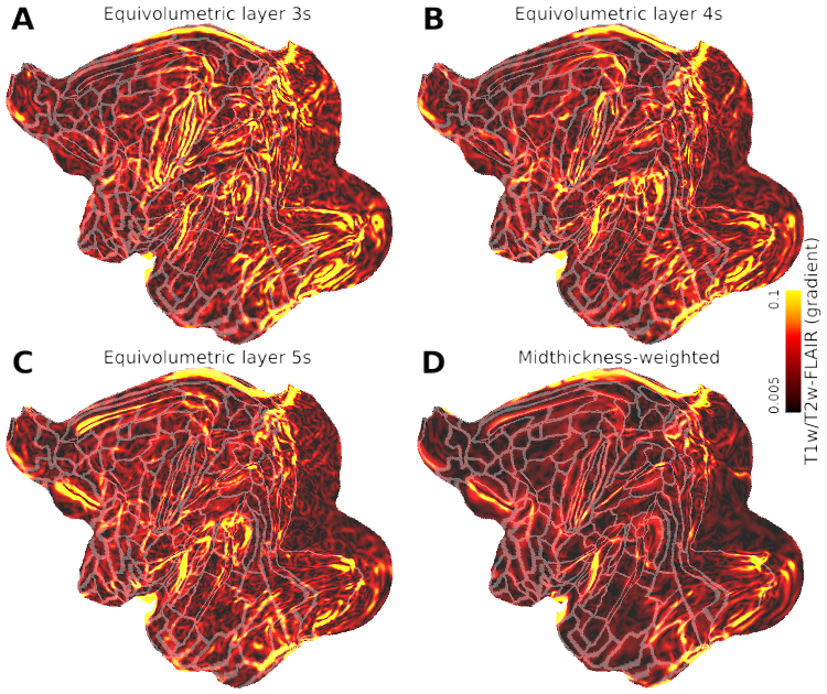
- Figure 4. Equivolumetric Layer-specific Myelin Transitions provide Improved Sensitivity to Cortical Area Boundaries Compared to Midthickness-weighted Gradients.
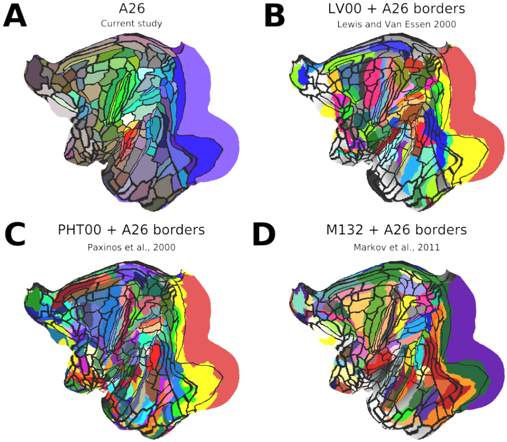
- Figure 5. Comparison of Cortical Area Atlases from Four Studies.
- Figure 7A, C. Comparison of macaque and human parcellation of cerebral cortex.
- Supplementary Figure 7. Charting Bands of Baillarger in the macaque cerebral cortex.
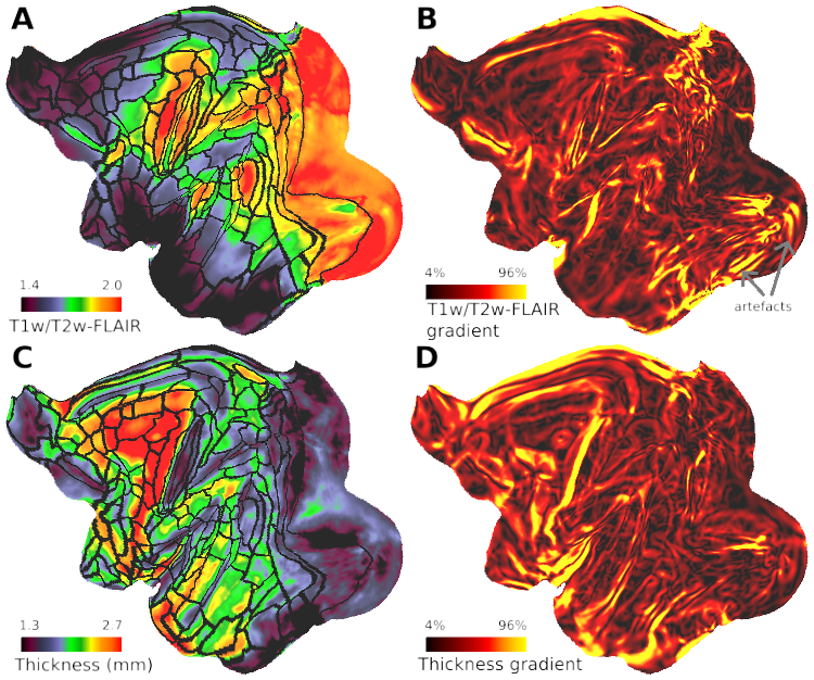
- Supplementary Figure 8. Comparison of equivolumetric, midthickness-weighted and cortical thickness gradients.
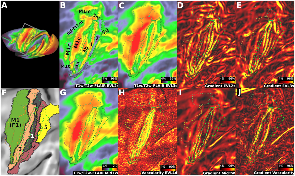
- Supplementary Figure 9. Charting Area Boundaries and Areal Subdivisions in the Sensorimotor Cortices.
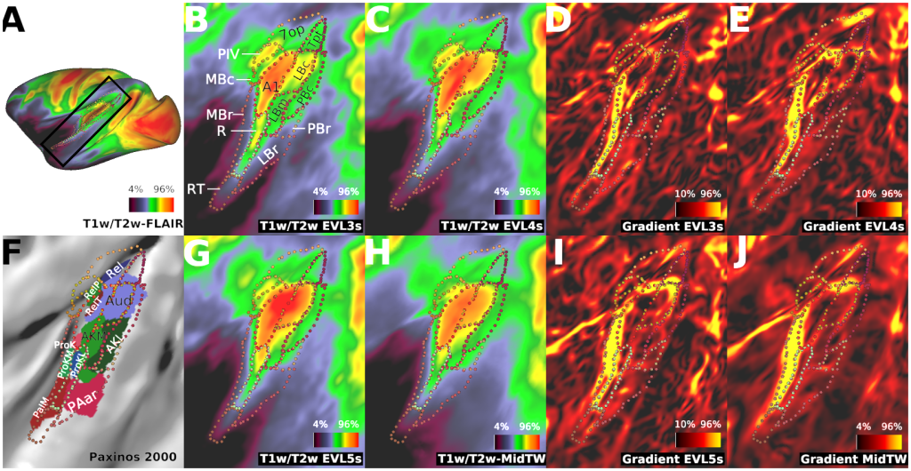
- Supplementary Figure 10. Charting Area Boundaries in the Auditory and Vestibular Cortex.
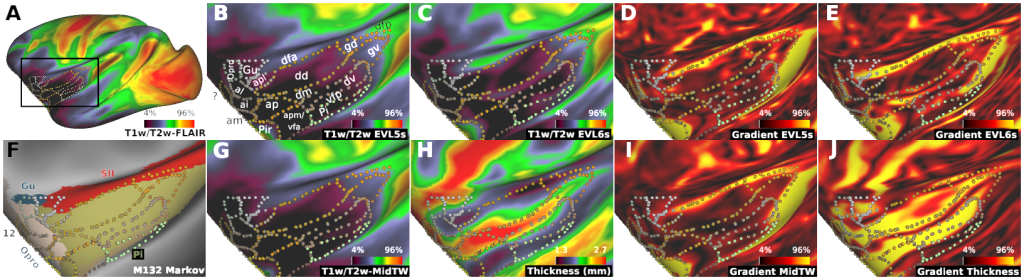
- Supplementary Figure 11. Charting Area Boundaries in Insular Cortex.
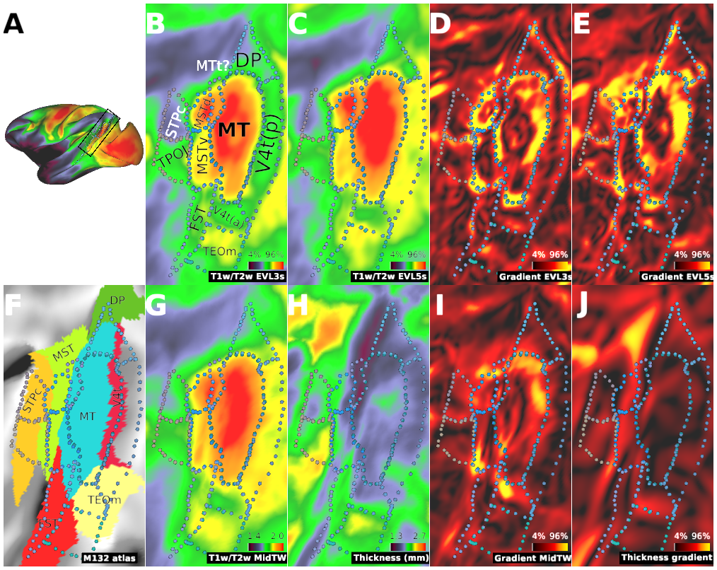
- Supplementary Figure 12. Charting Area Boundaries adjacent to MT complex (MT+).
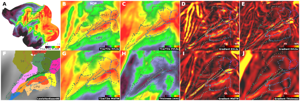
- Supplementary Figure 13. Charting Area Boundaries in Intraparietal Cortex.
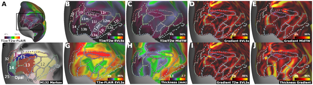
- Supplementary Figure 14. Charting Area Boundaries in Orbitofrontal Cortex.