
FULL TITLE:
The spatial extent of anatomical connections within the thalamus varies across the cortical hierarchy in humans and macaques
SPECIES:
Macaque, Human
DESCRIPTION:
Much of this work was made possible by the commitment of scientists who openly share their data. To acknowledge that effort, please credit the original authors when using these files.
These files include those from:
- Human Connectome Project MRI data - Minimally preprocessed diffusion, structural, and functional MRI from the Human Connectome Project (Van Essen et al., 2013, NeuroImage; https://doi.org/10.1523/JNEUROSCI.0493-16.2016)
- Macaque tractography datasets - Warrington et al., 2022, Science Advances (https://doi.org/10.1126/sciadv.abq2022); Mars et al., 2018, eLife (https://doi.org/10.7554/eLife.35237); Folloni et al., 2019, eLife (https://doi.org/10.7554/eLife.47175)
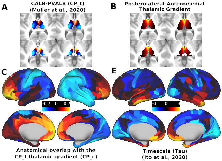
- Cortical timescale map - Ito et al., 2020, NeuroImage (https://github.com/ColeLab/hierarchy2020; https://doi.org/10.1016/j.neuroimage.2020.117141)
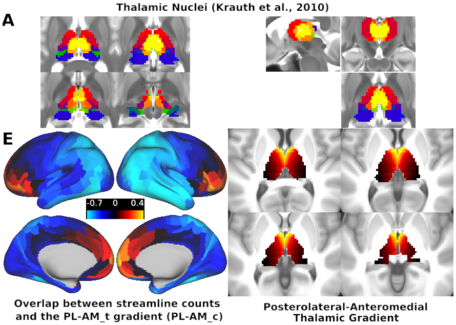
- Thalamic nuclei ROIs - Originally obtained in 1 mm space from Krauth et al., 2010, NeuroImage (https://doi.org/10.1016/j.neuroimage.2009.10.042) and subsequently downsampled to 2 mm MNI space.
- Thalamic core–matrix atlas - Müller et al., 2020, NeuroImage (https://github.com/macshine/corematrix)
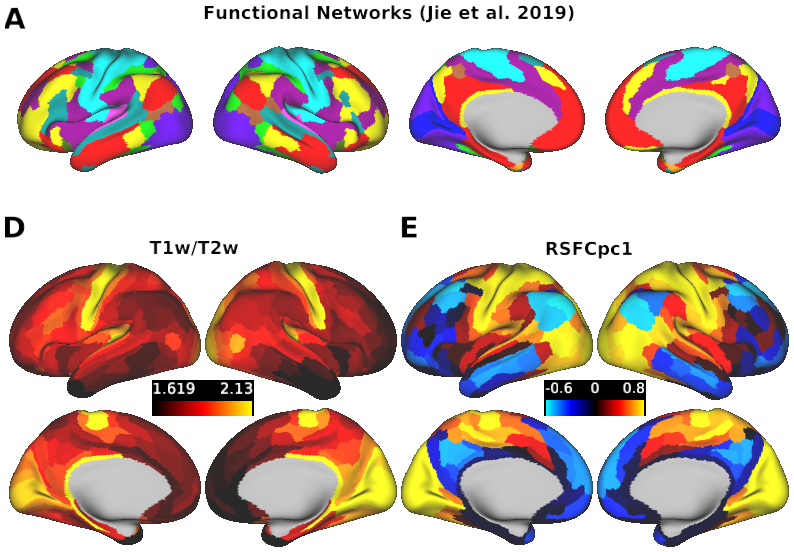
- Cole–Anticevic Brain-wide Network Partition (CAB‑NP) - Ji, Spronk et al., 2019, NeuroImage (https://github.com/ColeLab/ColeAnticevicNetPartition; https://doi.org/10.1016/j.neuroimage.2018.10.006; https://balsa.wustl.edu/study/show/wZML)
- Glasser HCP-MMP1.0 Parcellation - Glasser et al., 2016, Nature (https://doi.org/10.1038/nature18933)
- Melbourne Subcortical Atlas - Tian et al., 2020, Nature Neuroscience (https://doi.org/10.1038/s41593-020-00711-6; https://github.com/yetianmed/subcortex)
- Yeo Atlas - Yeo et al., 2011 J Neurophysiol (https://doi.org/10.1152/jn.00338.2011)
- Macaque T1w/T2w maps - Hayashi et al., 2021, NeuroImage (https://doi.org/10.1016/j.neuroimage.2021.117726; https://balsa.wustl.edu/study/Klr0B)
- Macaque parcellation - Van Essen et al., 2019, PNAS (https://doi.org/10.1073/pnas.1902299116; https://balsa.wustl.edu/study/976M4), based on the Markov atlas (Markov et al., 2014, Cerebral Cortex; https://doi.org/10.1093/cercor/bhs270)
ABSTRACT:
Each cortical area has a distinct pattern of anatomical connections within the thalamus, a central subcortical structure composed of functionally and structurally distinct nuclei. Previous studies have suggested that certain cortical areas may have more extensive anatomical connections that target multiple thalamic nuclei, which potentially allows them to modulate distributed information flow. However, there is a lack of quantitative investigations into anatomical connectivity patterns within the thalamus. Consequently, it remains unknown if cortical areas exhibit systematic differences in the extent of their anatomical connections within the thalamus. To address this knowledge gap, we used diffusion magnetic resonance imaging (dMRI) to perform brain-wide probabilistic tractography for 828 healthy adults from the Human Connectome Project. We then developed a framework to quantify the spatial extent of each cortical area’s anatomical connections within the thalamus. Additionally, we leveraged resting-state functional MRI, cortical myelin, and human neural gene expression data to test if the extent of anatomical connections within the thalamus varied along the cortical hierarchy, from sensory and motor to multimodal associative cortical areas. Our results revealed two distinct corticothalamic tractography motifs: 1) a sensorimotor cortical motif characterized by focal thalamic connections targeting posterolateral thalamus, associated with fast, feed-forward information flow; and 2) an associative cortical motif characterized by diffuse thalamic connections targeting anteromedial thalamus, associated with slow, feed-back information flow. These findings were consistent across human subjects and were also observed in macaques, indicating cross-species generalizability. Overall, our study demonstrates that sensorimotor and association cortical areas exhibit differences in the spatial extent of their anatomical connections within the thalamus, which may support functionally-distinct cortico-thalamic information flow.
PUBLICATION:
eLife
- DOI:
10.7554/eLife.95018.2.sa4
- Amber M Howell
- Shaun Warrington
- Clara Fonteneau
- Youngsun T Cho
- Stamatios N Sotiropoulos
- John D Murray
- Alan Anticevic
- University of Nottingham
- Yale University
-
CorticothalamicConnectivityPatterns_Figures.scene
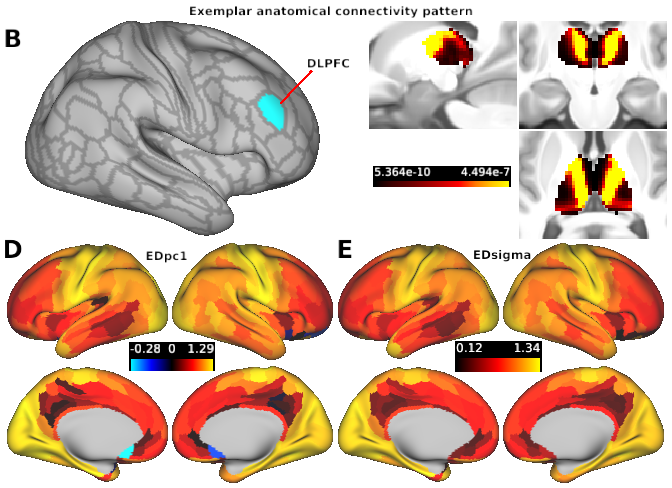
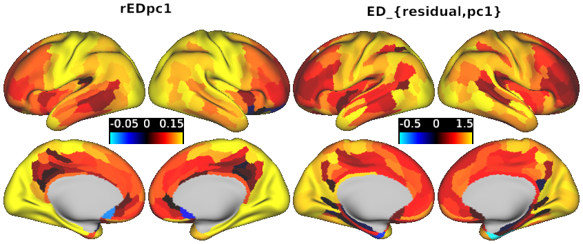
SCENES:- Fig. 1 - Workflow for quantifying the spread of cortical parcel's thalamic anatomical connectivity patterns using Euclidean distance (ED)
- Fig. 2 - Differences in thalamic connectivity patterns between sensory and association cortical parcels
- Figure 3 Mapping between the spatial extent of anatomical connections within the thalamus and anatomical coupling with the anteromedial-posterolateral thalamic gradient.
- Fig 4 Mapping between cortical variation in the extent of thalamic connectivity patterns, anatomical overlap across thalamic subpopulations, and intrinsic timescale.
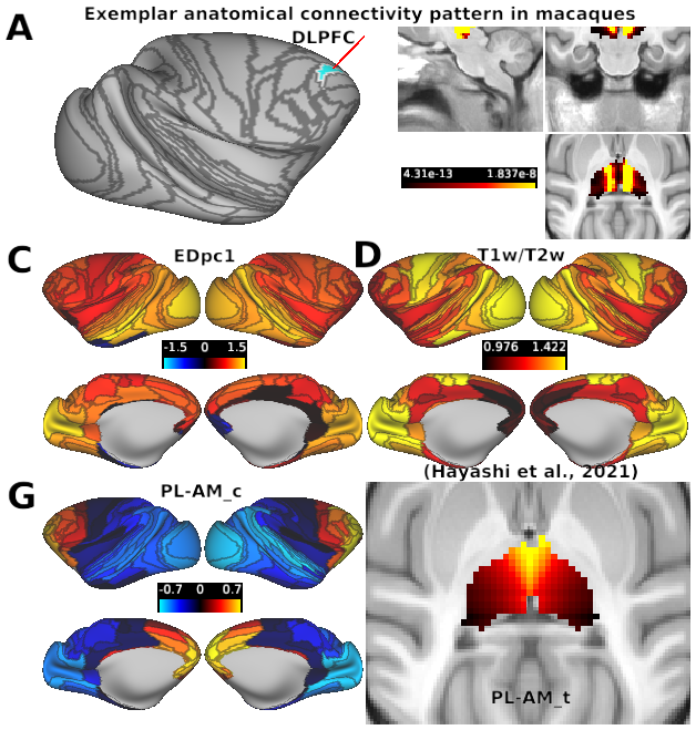
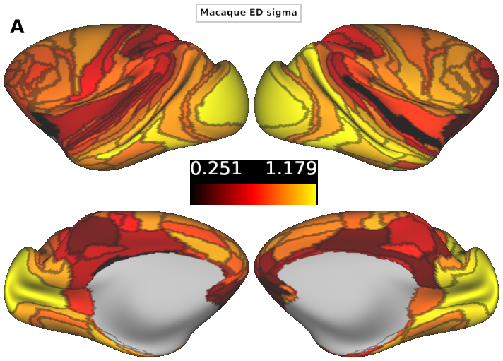
- Fig 5 Cortical variation of the extent of anatomical connections within the macaque thalamus.
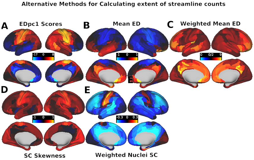
- S4 - Alternative methods for calculating spatial extent of streamline counts
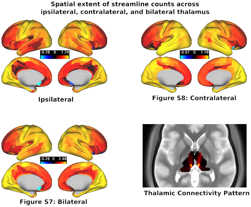
- S7-8. Laterality comparison
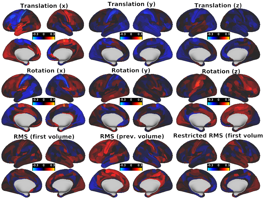
- Fig. S9 Motion Controls
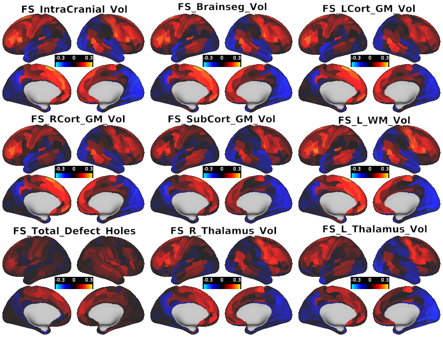
- Figure S10 - Freesurfer Controls
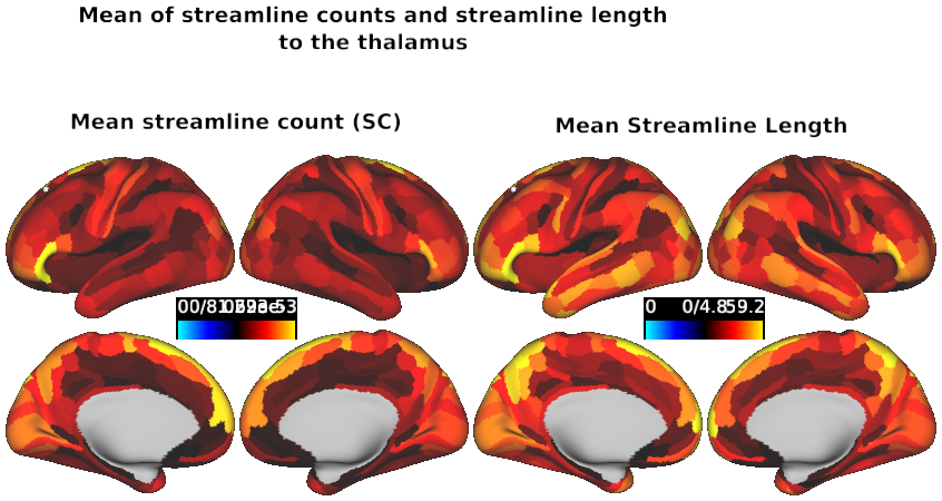
- S11 Mean Streamline Count Comparison
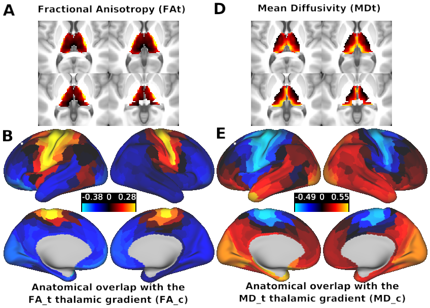
- Figure S12 - FA and MD
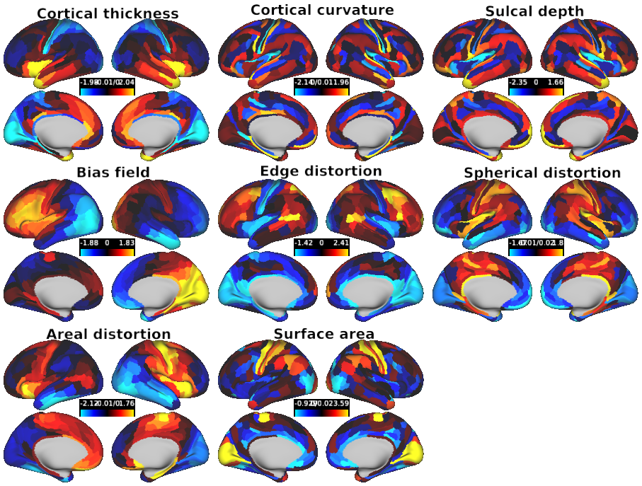
- Figure S13 -Cortical geometry controls
- Figure S14 S15
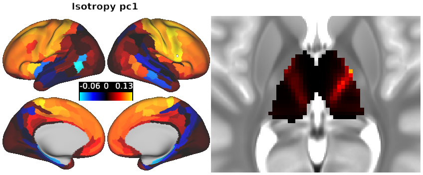
- Figure S16 - Isotropy
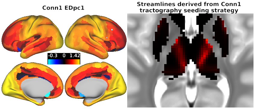
- Figure S19 - Conn1 Replication
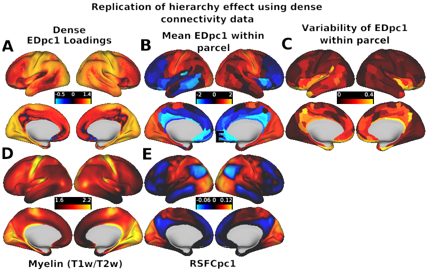
- S20 - Dense ED - Figure S20 - Dense ED
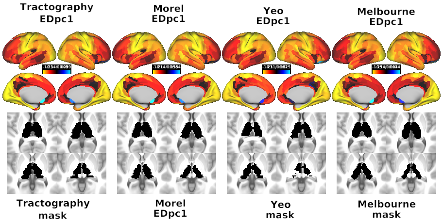
- S21 Thalamic Parcellation comparisons - Figure S21 - Thalamic ROI Comparisons
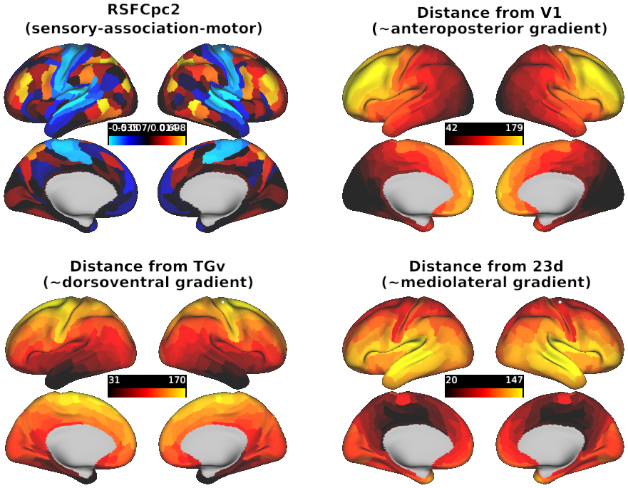
- Figure S22 Hierarchical Gradient Specificty
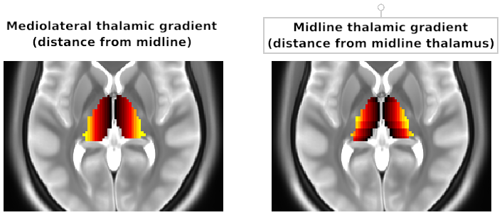
- Figure S26 - Comparison of medioventral and midline thalamus gradients
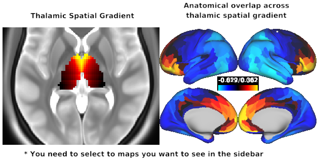
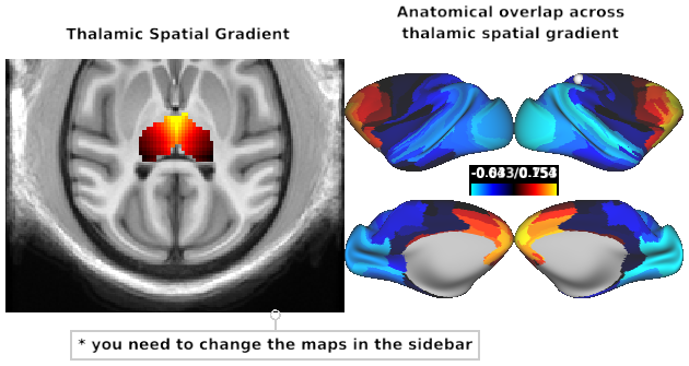
- Figure S27 - Human Thalamic Spatial Gradient Overlap
- Fig S29 Comparisons between human and macaque cortical connectivity patterns
- Figure S30 - Macaque Thalamic Spatial Gradient Overlap
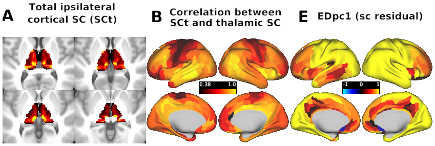
- S31 Correspondence between group-level EDpc1 loadings before and after regressing out each thalamic voxel’s total streamline count to all ipsilateral cortical regions (SCt).