FULL TITLE:
A 2020 View of Tension-Based Cortical Morphogenesis
SPECIES:
Human
DESCRIPTION:
The scenes for this study were created using a newer version of wb_view, they will not display properly using wb_view v1.4.2. Please keep an eye out for the new wb_view release coming early 2021.
ABSTRACT:
Mechanical tension along the length of axons, dendrites, and glial processes has been proposed as a major contributor to morphogenesis throughout the nervous system (Van Essen, D.C., Nature 385: 313-318, 1997). Tension-based morphogenesis (TBM) is a conceptually simple and general hypothesis based on physical forces that help shape all living things. Moreover, if each axon and dendrite strives to shorten while preserving connectivity, aggregate wiring length would remain low. TBM can explain key aspects of how the cerebral and cerebellar cortices remain thin, expand in surface area, and acquire their distinctive folds. This article reviews progress since 1997 relevant to TBM and other candidate morphogenetic mechanisms. At a cellular level, studies of diverse cell types in vitro and in vivo demonstrate that tension plays a major role in many developmental events. At a tissue level, I propose a Differential Expansion Sandwich Plus (DES+) revision to the original TBM model for cerebral cortical expansion and folding. It invokes tangential tension and “sulcal zipping” forces along the outer cortical margin as well as tension in the white matter core, together competing against radially biased tension in the cortical gray matter. Evidence for and against the DES+ model is discussed, and experiments are proposed to address key tenets of the DES+ model. For cerebellar cortex, a Cerebellar Multi-layer Sandwich (CMS) model is proposed that can account for many distinctive features, including its unique, accordion-like folding in the adult, and experiments are proposed to address its specific tenets.
PUBLICATION:
Proceedings of the National Academy of Sciences of the United States of America
- DOI:
10.1073/pnas.2016830117
- David C Van Essen
- Washington University in St. Louis
-
VanEssen_TBM_PNAS2020_Caudate_FigS2.scene
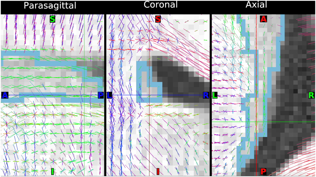
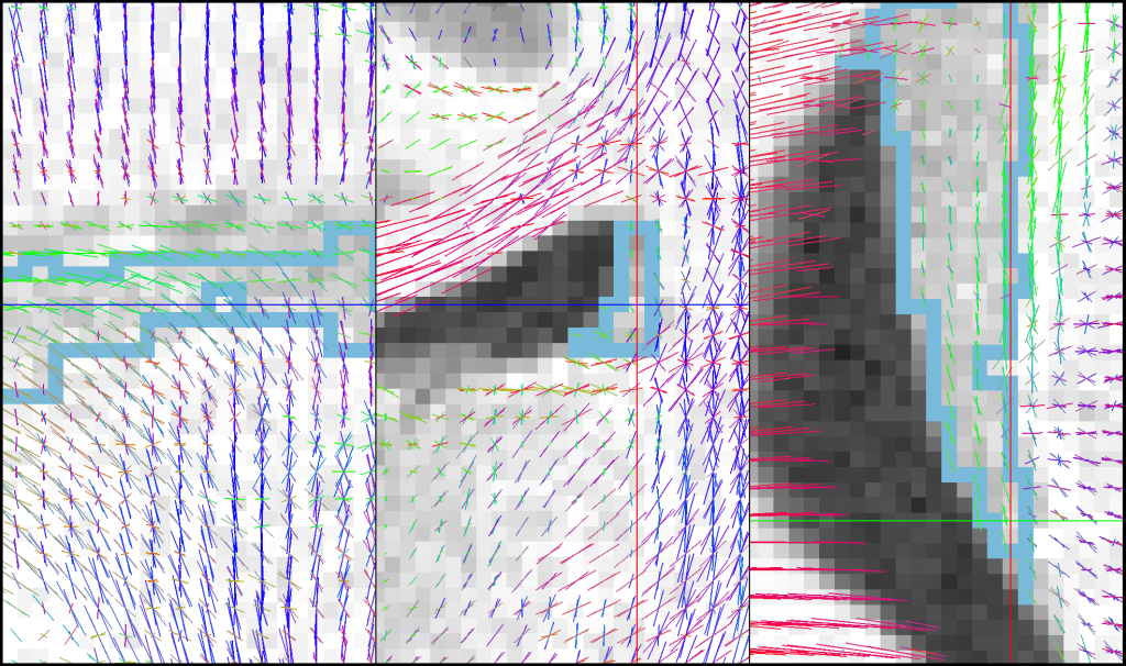
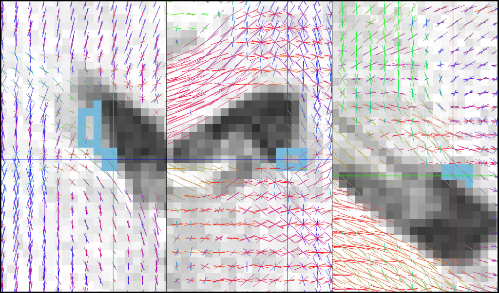
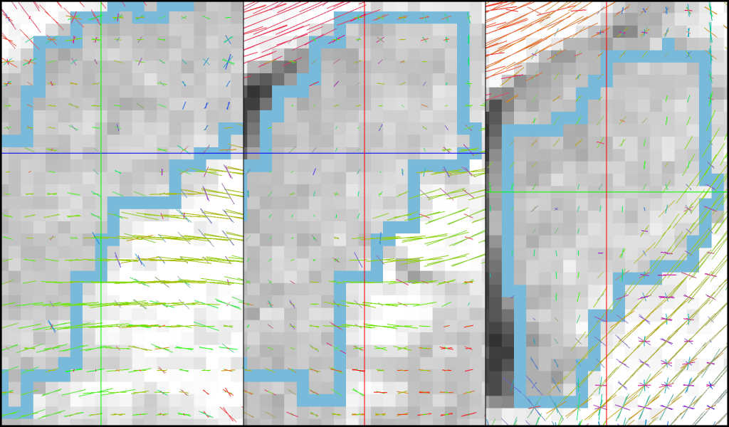
SCENES:- Figure S2A - Fiber bundle orientation in human caudate nucleus, anterior tail
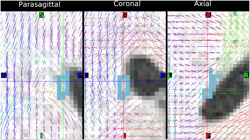
- Figure S2B - Fiber bundle orientation in human caudate nucleus oblique posterior tail
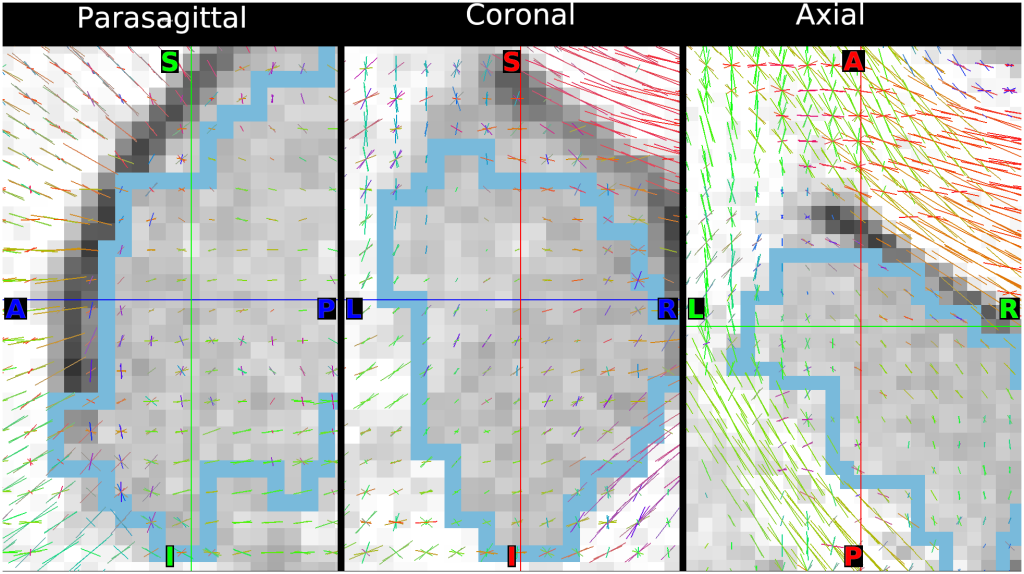
- Figure S2C - Fiber bundle orientation in human head of caudate nucleus
- 100307 anterior tail of Caudate - Right hemisphere
- 100307 oblique posterior tail of Caudate - Right hemisphere
- 100307 Head of Caudate - Right hemisphere

