
Scene Preview
study:
Tension-based Morphogenesis
SCENE FILE:
VanEssen_TBM_PNAS2020_Caudate_FigS2
SCENE:
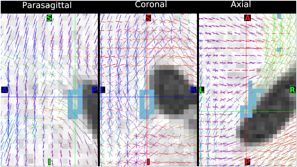
Figure S2B - Fiber bundle orientation in human caudate nucleus oblique posterior tail
DESCRIPTION:
100307 Left Posterior Tail Oblique - centered at 155,140,124
Fig. S2. Fiber bundle orientation in and near human caudate nucleus revealed by diffusion MRI from the Human Connectome Project (HCP). Data from exemplar HCP subject 100307 parasagittal (left), coronal (middle) and axial (right) panels from the anterior portion of the caudate tail (row A), a more posterior region (row B), and the head of the caudate (row C). Diffusion imaging scans acquired with 1.25 mm voxel size and other imaging parameters described in ref. [15]
TAGS:
Modality:T1-weighted, Modality:T2-weighted, Modality:Myelin Map, Registration:MSMAll, Species:Human