
Scene Preview
study:
Parcellating Cerebral Cortex
SCENE FILE:
VanEssen_Glasser_2018_NeuronReview_SI_Fig2_Retinotopy
SCENE:
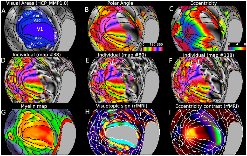
SI Figure 2 Human visuotopic organization
DESCRIPTION:
SI Figure 2. Panel A: early visual areas in the HCP_MMP1.0 parcellation. Note that the adjacency of ventral V2 to ProS, PreS, and PHA1 (but not consistently for the 210P vs 210V group average parcellations). Panel D: map 38; subject 157336. Panel E: map 80; subject 198653. Panel F: map 138; subject 644246. Note that SNR (R2 maps) were robust in V1 – V3 in all three subjects, even in regions showing atypical retinotopy.
TAGS:
Surface Mesh:32k fs LR, Registration:MSMAll, Modality:Myelin Map, Species:Human, Parcellation:HCP_MMP1.0