
Scene Preview
study:
Anatomical variability, coordinates and targeting in marmoset brain
SCENE FILE:
MarmosetRIKEN20MultimodalTemplate
SCENE:
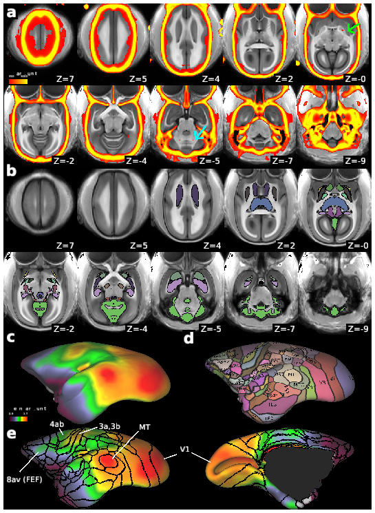
Figure 8. The MarmosetRIKEN20 multi-modal templates in AC-PC and grayordinates
DESCRIPTION:
(a) The multi-modal templates with T1w MRI (grayscale, N = 20) overlaid with thresholded CT (red-yellow, N = 10). Each subject’s CT image was registered to T1w MRI in AC-PC native coordinates using the MBFR+BBR, warped to the AC-PC template coordinates using an MRI warp field, and averaged across subjects. Note that physiological calcifications were found bilaterally in the globus pallidus (green arrow, z = 0) and dentate nucleus (cyan arrow, z = -5). (b) the subcortical gray matter atlas of MarmosetRIKEN20 including 21 subcortical gray matter regions, and anterior and posterior commissures (color coded, outlined by black line) overlaid on the T1w-weighted template image (gray color). (c) T1w divided by T2w myelin map in color overlaid on the average midthickness surfaces of MarmosetRIKEN20 grayordinates. (d) Surface version of marmoset cortical parcellation atlas of Paxinos, Watson, Petrides, Rosa and Tokuno (Paxinos et al., 2012) including 116 cortical areas. (e) Outlines of the cortical parcellations overlaid on the myelin map (left, lateral; right, medial view). Note that high myelin contrast is colocalized with the parcellations at MT, somatomotor sensory areas (4ab, 3a, 3b) and visual cortex (V1), and weakly high myelin with the area 8ab, frontal eye field (FEF).
TAGS: