
There is also a preprint version of this study here.
FULL TITLE:
Anatomical variability, multi-modal coordinate systems, and precision targeting in the marmoset brain
SPECIES:
Marmoset
ABSTRACT:
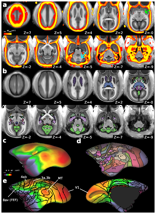
Localising accurate brain regions needs careful evaluation in each experimental species due to their individual variability. However, the function and connectivity of brain areas is commonly studied using a single-subject cranial landmark-based stereotactic atlas in animal neuroscience. Here, we address this issue in a small primate, the common marmoset, which is increasingly widely used in systems neuroscience. We developed a non-invasive multi-modal neuroimaging-based targeting pipeline, which accounts for intersubject anatomical variability in cranial and cortical landmarks in marmosets. This methodology allowed creation of multi-modal templates (MarmosetRIKEN20) including head CT and brain MR images, embedded in coordinate systems of anterior and posterior commissures (AC-PC) and CIFTI grayordinates. We found that the horizontal plane of the stereotactic coordinate was significantly rotated in pitch relative to the AC-PC coordinate system (10 degrees, frontal downwards), and had a significant bias and uncertainty due to positioning procedures. We also found that many common cranial and brain landmarks (e.g., bregma, intraparietal sulcus) vary in location across subjects and are substantial relative to average marmoset cortical area dimensions. Combining the neuroimaging-based targeting pipeline with robot-guided surgery enabled proof-of-concept targeting of deep brain structures with an accuracy of 0.2 mm. Altogether, our findings demonstrate substantial intersubject variability in marmoset brain and cranial landmarks, implying that subject-specific neuroimaging-based localization is needed for precision targeting in marmosets. The population-based templates and atlases in grayordinates, created for the first time in marmoset monkeys, should help bridging between macroscale and microscale analyses.
PUBLICATION:
NeuroImage
- DOI:
10.1016/j.neuroimage.2022.118965
- Takayuki Ose
- Joonas A. Autio
- Masahiro Ohno
- Stephen Frey
- Akiko Uematsu
- Akihiro Kawasaki
- Chiho Takeda
- Yuki Hori
- Kantaro Nishigori
- Tomokazu Nakako
- Chihiro Yokoyama
- Hidetaka Nagata
- Tetsuo Yamamori
- David Van Essen
- Matthew F. Glasser
- Hiroshi Watabe
- Takuya Hayashi
- Rogue Research
- Sumitomo Dainippon Pharma Co., Ltd
- Kyoto University
- Washington University in St. Louis
- RIKEN
- Tohoku University