
Scene Preview
study:
Cortical-subcortical functional networks
SCENE FILE:
ColeAnticevicNetworkPartition_MainFigures
SCENE:
Figure 5
DESCRIPTION:
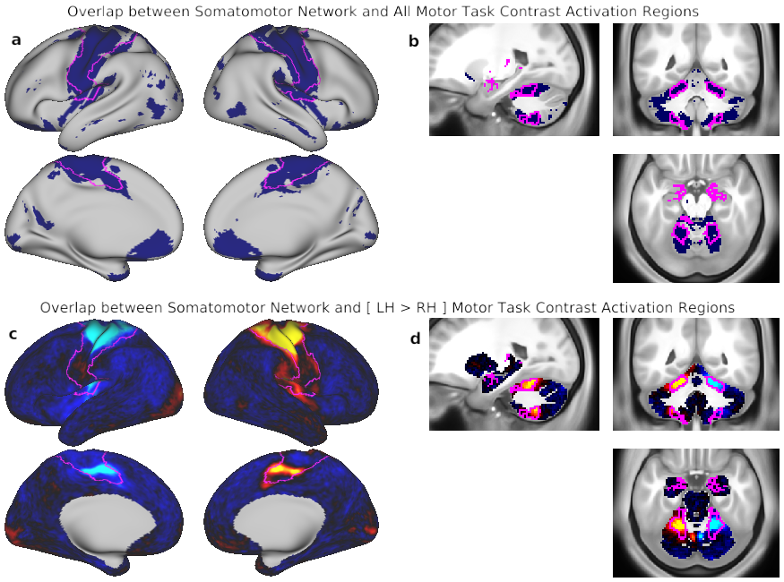
Fig. 5. Convergence of cortical and subcortical network partition and motor task activation. The motor network is shown as evidence for valid extension of cortical functional networks to subcortical regions. A) Combined motor task responses for comparisons between two movements [(left foot > tongue), (left hand > tongue), (right hand > tongue), (right foot > tongue), and (tongue > right foot)] in the cortex, with the SMN outlined in fuchsia. B) Combined motor task responses in the subcortex, with the SMN from the wGSR subcortical parcellation outlined in fuchsia. For ease of comparison, SMN from the woGSR parcellation is also loaded and can be toggled into display. Because the degree of convergence with task activation is higher for the wGSR than the woGSR version, we use the wGSR parcellation for all subsequent subcortical analyses presented in this study. C) Map of the left foot (LH) > right hand (RH) contrast in the cortex, with the SMN outlined in green. D) Map of the left hand (LH) > right hand (RH) contrast in the subcortex, with the SMN from the wGSR subcortical parcellation outlined in fuchsia. The SMN from the woGSR subcortical parcellation is loaded and can be toggled into display. Note the ipsilateral representation of the hand movements in the cerebellum and the higher convergence of the wGSR parcels relative to the woGSR parcels with task activation.
TAGS:
Surface Mesh:32k fs LR, Modality:T1-weighted, Species:Human