
Scene Preview
study:
Mapping the human corticoreticular pathway
SCENE FILE:
Figures
SCENE:
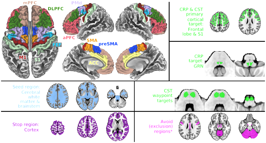
Figure 2. Tractography regions
DESCRIPTION:
Top left panel: Frontal cortex and S1 regions of interest on the gray/white matter boundary surface. This region was mapped to volume space and dilated 2 mm inward for use as the primary cortical region of interest during tractography (top right panel). Remaining panels: streamline inclusion regions (lime green), seeding region (pale blue), stopping region (purple) and exclusion regions (fuscia), in the MNI tractography space (MNI152NLin2009cAsym). *Exclusion regions were: corpus callosum, anterior commissure, cortical origin of corticobulbar fibers, superior medial thalamus, cranial nerves II
TAGS:
Species:Human