
Scene Preview
study:
The Impact of Traditional Neuroimaging Methods on the Spatial Localization of Cortical Areas
SCENE FILE:
Coalson_et_al_2018
SCENE:
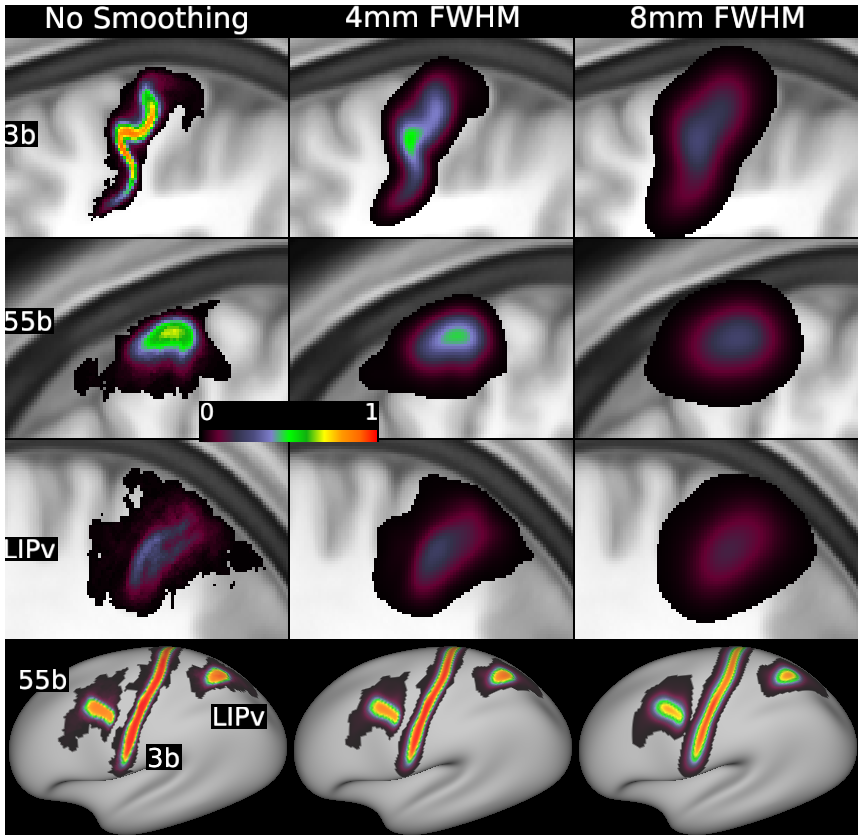
Figure 4
DESCRIPTION:
Effects of volume-based and surface-based smoothing on example cortical areas. The Top three rows show enlarged sagittal slices of volumetric probabilistic maps through the maximum probability of three exemplar areas, before (Left) and after unconstrained volume-based Gaussian smoothing of 4-mm (Center) or 8-mm (Right) FWHM. In each row, white dots are in corresponding positions for reference. The Bottom row shows the same amounts of surface-based Gaussian smoothing applied to the same three areas after areal-feature–based registration (MSMAll). Areal probability values decrease in the volume after smoothing substantially more than on the surface with the same amount in millimeters FWHM of smoothing. See SI Appendix, Supplemental Methods M3 and M4.
TAGS:
Registration:MSMAll, Surface Mesh:32k fs LR, Modality:T1-weighted