
Scene Preview
study:
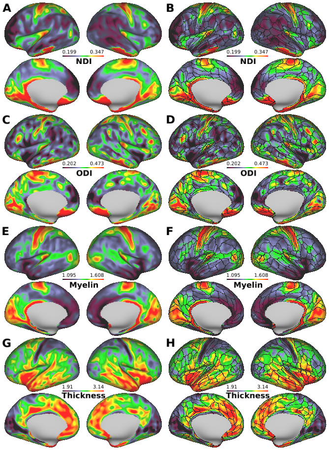
Neurite imaging reveals microstructural variations in human cerebral cortical gray matter.
SCENE FILE:
Figures_NODDI
SCENE:
FigS7
DESCRIPTION:
Single-subject's orientation dispersion index (ODI) and neurite density index (NDI) cortical maps from the Human Connectome Project (HCP) data. The surface metrics have been smoothed with 5 mm FWHM surface geodesic Gaussian smoothing. (A) Neurite density index (NDI), (C) orientation dispersion index (ODI), (E) myelin and (G) cortical thickness. (B, D, F and H) The imaging modalities superimposed with the boundaries of the Human Connectome Project (HCP) multi-modal parcellations (black lines). This representative single-subject shows similar cortical distribution patterns of ODI and NDI compared with 505-subject average maps (see Fig. 1).
TAGS:
Surface Mesh:32k fs LR, Registration:MSMAll, Modality:Myelin Map, Species:Human