
Scene Preview
study:
Neurite imaging reveals microstructural variations in human cerebral cortical gray matter.
SCENE FILE:
Figures_NODDI
SCENE:
Fig2
DESCRIPTION:
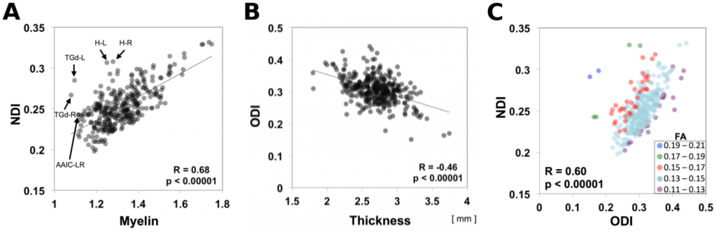
Correlation between neurite orientation dispersion and density imaging (NODDI) and cortical architectural properties for each of the HCP_MMP1.0 areas. (A) Neurite density index (NDI) plotted against myelin. (B) Orientation dispersion index (ODI) plotted against cortical thickness. (C) NDI plotted against ODI classified by the value of FA. Each data point represents 505-subject mean values for each of the 300-parcels, where SNR exceeded 17. Abbreviations: H-L: left hippocampus; H-R: right hippocampus; TGd-L: left area TG dorsal; TGd-R: right area TG dorsal; AAIC-LR: left and right anterior agranular insula complex.
TAGS:
Surface Mesh:164k fs LR, Registration:MSMAll, Parcellation:HCP_MMP1.0