
Scene Preview
study:
Dynamic Patterns of Cortical Expansion in Preterm Human Development
SCENE FILE:
DynamicPatternsHumanPretermExpansion
SCENE:
Figure4
DESCRIPTION:
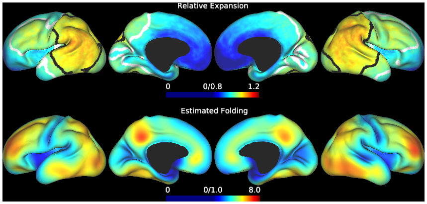
Fig. 4(B). Gradients of cortical expansion and folding are consistent across subjects. For the period from 30 to 38 weeks PMA, relative area expansion (top) and increased regional folding (bottom) were highest in the lateral parietal-temporal-occipital region and lateral frontal lobe (n=20). Black and white contours enclose regions where relative expansion is significantly higher and lower, respectively, than the global average. Individual maps are also available for viewing within this scene file:
Relative expansion, left = PMA30to38.L.noninjured20.average.GGnorm.func.gii
Relative expansion, right = PMA30to38.R.noninjured20.average.GGnorm.func.gii
Estimated folding, left = PMA30to38.L.noninjured20.AbsNormMeanCurvSmoothed4Diff.func.gii
Estimated folding, right = PMA30to38.R.noninjured20.AbsNormMeanCurvSmoothed4Diff.func.gii
TAGS:
Species:Human