


FULL TITLE:
Functionally and structurally distinct fusiform face area(s) in over 1000 participants
SPECIES:
Human
DESCRIPTION:
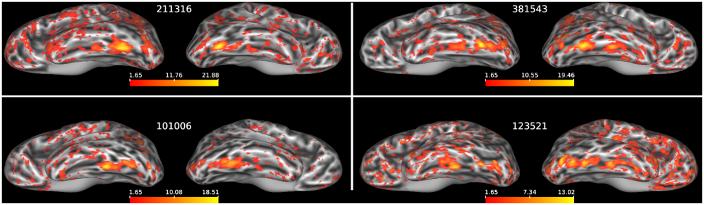
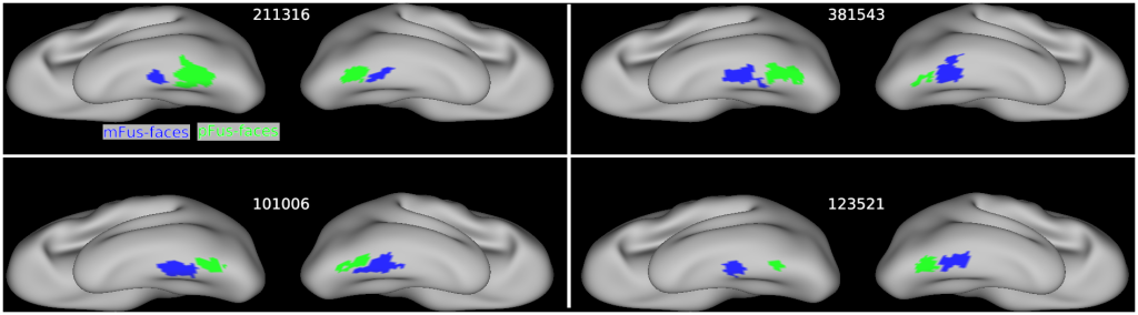
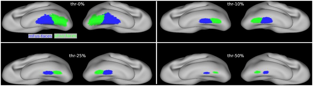
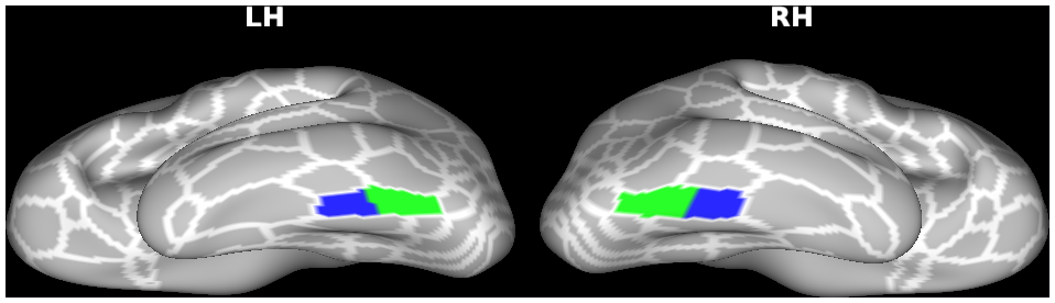
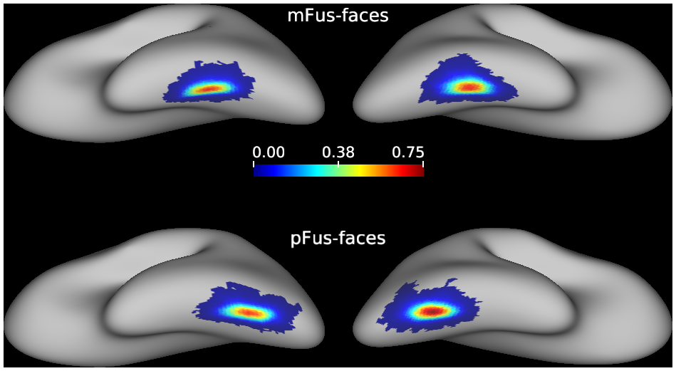
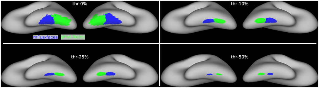
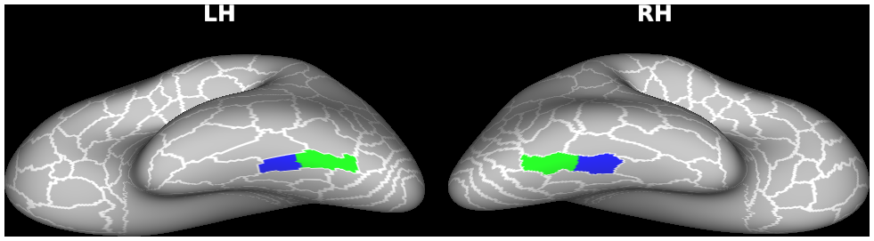
Face-selective regions on the fusiform gyrus (FG), or "Fusiform Face Areas (FFAs)", were defined as either mFus-faces/FFA-2 or pFus-faces/FFA-1 for each of 1053 participants from Human Connectome Project Young Adult (HCP-YA, S1200 data release, 2017) by taking both individual cortical landmarks (OTS: occipito-temporal sulcus; CoS: collateral sulcus; MFS: mid-fusiform sulcus) and face-selective activation clusters (faces versus others, Z>1.65, p<0.05, uncorrected) into account. At least one face-selective region was identifiable in every hemisphere in each participant and 95.44% of hemispheres had two face-selective regions on the FG. The probabilistic maps and maximum probability maps (MPMs) were also created for the face-selective regions. All maps are provided in both 32k_fs_LR and fsaverage spaces.
Please see readme.pdf for more information about this dataset.
ABSTRACT:
The fusiform face area (FFA) is a widely studied region causally involved in face perception. Even though cognitive neuroscientists have been studying the FFA for over two decades, answers to foundational questions regarding the function, architecture, and connectivity of the FFA from a large (N>1000) group of participants are still lacking. To fill this gap, we quantified these features of fusiform face-selective regions in 1053 participants in the Human Connectome Project. After manually defining over 4,000 fusiform face-selective regions, we report five main findings. First, 68.76% of hemispheres have two cortically separate regions (pFus-faces/FFA-1 and mFus-faces/FFA-2). Second, in 26.69% of hemispheres, pFus-faces/FFA-1 and mFus-faces/FFA-2 are spatially contiguous, yet functionally, architecturally, and connectivity distinct. Third, pFus-faces/FFA-1 is more face-selective than mFus-faces/FFA-2, and the two regions have distinct functional connectivity fingerprints. Fourth, pFus-faces/FFA-1 is cortically thinner and more heavily myelinated than mFus-faces/FFA-2. Fifth, face-selective patterns and functional connectivity fingerprints of each region are more similar in monozygotic than dizygotic twins and more so than architectural gradients. As we share our areal definitions with the field, future studies can explore how structural and functional features of these regions will inform theories regarding how visual categories are represented in the brain.
PUBLICATION:
BioRxiv
- DOI:
10.1101/2022.04.08.487562
- Xiayu Chen
- Xingyu Liu
- Benjamin J. Parker
- Zonglei Zhen
- Kevin S. Weiner
- State Key Laboratory of Cognitive Neuroscience and Learning, Beijing Normal University, Beijing 100875, China
- Department of Psychology, University of California, Berkeley, CA 94720
- Helen Wills Neuroscience Institute, University of California, Berkeley, CA 94720