


FULL TITLE:
A whole-brain 3D myeloarchitectonic atlas: mapping the Vogt-Vogt legacy to the cortical surface
SPECIES:
Human
DESCRIPTION:
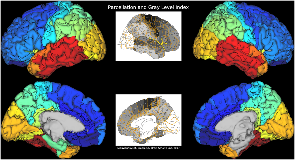
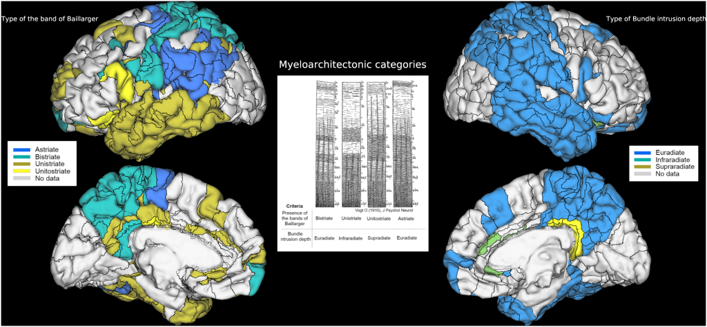
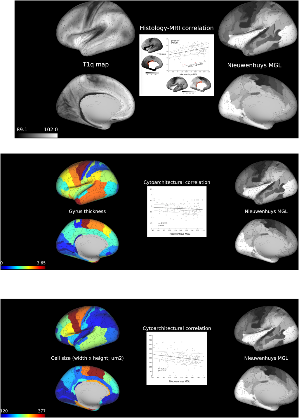
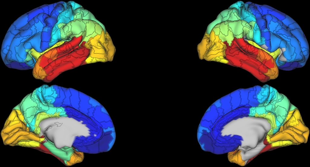
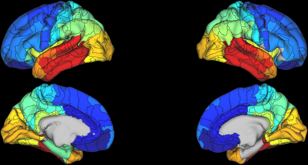
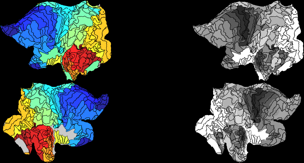
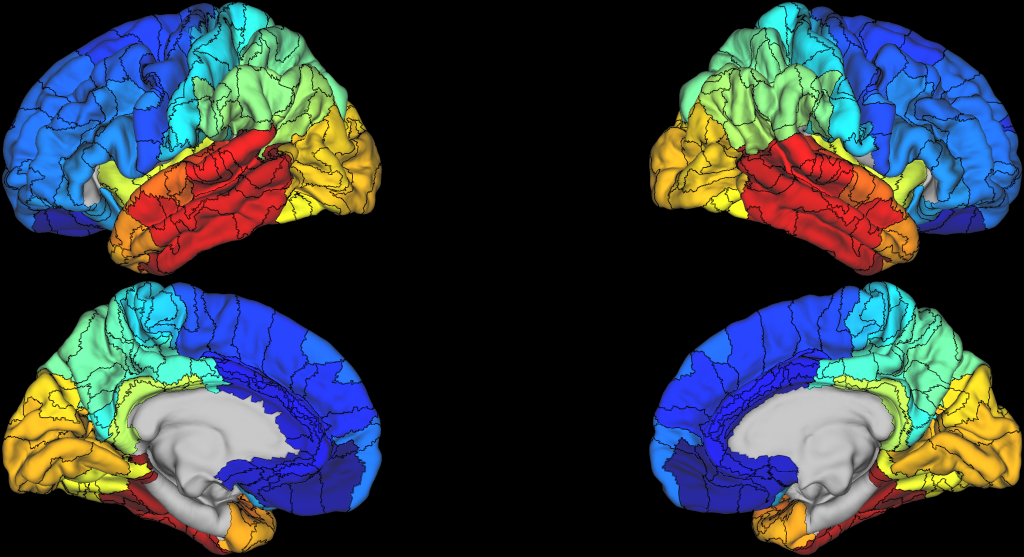
Here we share a) ready-to-use myeloarchitectonic parcellations in common space aligned with several brain templates in both volumetric and surface formats, b) raw and processed histology slices used for constructing the atlas and their original publications, c) intracortical laminar profiles of myelin content derived from photometric data of Vogt and Hopf, and, finally, d) Bash shell and MATLAB scripts to apply myeloarchitectonic parcellations to new projects.
ABSTRACT:
Building precise and detailed parcellations of anatomically and functionally distinct brain areas has been a major focus in Neuroscience. Pioneer anatomists parcellated the cortical manifold based on extensive histological studies of post-mortem brain, harnessing local variations in cortical cyto- and myeloarchitecture to define areal boundaries. Compared to the cytoarchitectonic field, where multiple neuroimaging studies have recently translated this old legacy data into useful analytical resources, myeloarchitectonics, which parcellate the cortex based on the organization of myelinated fibers, has received less attention. Here, we present the neocortical surface-based myeloarchitectonic atlas based on the histology-derived maps of the Vogt-Vogt school and its 2D translation by Nieuwenhuys. In addition to a myeloarchitectonic parcellation, our package includes intracortical laminar profiles of myelin content based on Vogt-Vogt-Hopf original publications. Histology-derived myelin density mapped on our atlas demonstrate close overlap with in vivo quantitative MRI markers for myelin and relates to cytoarchitectural features. Complementing the existing battery of approaches for digital cartography, the whole-brain myeloarchitectonic atlas offers an opportunity to validate imaging surrogate markers of myelin in both health and disease.
PUBLICATION:
NeuroImage
- DOI:
10.1016/j.neuroimage.2022.119617
- Foit, Niels Alexander
- Yung, Seles
- Lee, Hyo Min
- Bernasconi, Andrea
- Bernasconi, Neda
- Hong, Seok-Jun
- McGill University
-
COLIN27.scene
SCENES: -
MRI-Histology.scene
SCENES: -
conte69_10k.scene
SCENES: -
conte69_32k.scene
SCENES: -
fsaverage.scene
SCENES: -
fsaverage5.scene
SCENES: