
FULL TITLE:
Cortical Processing for the Vestibular and Visual Input of Egomotion in Macaque Monkeys: Separate Networks with Targeted Convergence
SPECIES:
Macaque
DESCRIPTION:
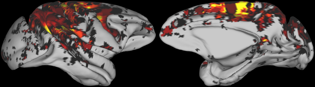
• NMT_rh.mid_surface.rsl.surf.gii:
Mid-thickness cortical surface of the right hemisphere of the macaque brain, derived from the NMT (National Institutes of Mental Health Macaque Template). This surface is used as the anatomical template onto which group-level functional data are projected.
• NMT_rh.mid_surface.rsl.GVS.func.gii:
Group-level functional map projected onto the right-hemisphere NMT mid-thickness surface, summarizing cortical activations elicited by galvanic vestibular stimulation (GVS). Values at each surface node indicate the number of hemispheres showing significant activation at that node, across the 8 hemispheres included in the study (4 macaque monkeys).
• NMT_rh.mid_surface.rsl.FLOW.func.gii:
Group-level functional map projected onto the same surface, summarizing cortical activations in response to visual optic flow stimulation. Values at each surface node indicate the number of hemispheres showing significant activation at that node, across the 8 hemispheres included in the study (4 macaque monkeys).
• NMT_rh.mid_surface.rsl.BOTHthr.func.gii:
Thresholded group-level functional map highlighting cortical regions showing overlapping activations in both the vestibular (GVS) and visual optic flow conditions.
ABSTRACT:
The integration of visual and vestibular inputs during egomotion is fundamental for both postural and navigational control. In the present study, we used functional magnetic resonance imaging (fMRI) in four macaque monkeys to investigate cortical activation in response to galvanic vestibular stimulation (GVS), applied through transmastoid electrodes, and egomotion-compatible (EC) optic flow patterns. Visual and vestibular stimulations activate two largely independent cortical networks: the vestibular network encompasses the insular cortex, superior parietal lobule, frontal lobe, and cingulate cortex, while optic flow primarily activates regions in the superior temporal sulcus, temporo-parietal junction, inferior parietal lobule, and restricted portions of the cingulate and frontal cortices. Despite this segregation, several areas exhibit visuo-vestibular convergence: VPS in the temporo-parietal junction, area 7 in the inferior parietal lobule, VIP and LIP in the intraparietal sulcus, MSTd in the superior temporal sulcus, CSv in the cingulate sulcus, and FEFsem in the frontal cortex. These findings demonstrate that visual and vestibular signals generated by egomotion are processed in extended and distinct cortical networks with several narrow convergence sites, consistent with the idea that multisensory integration during self-motion is achieved through selective convergence rather than general network overlap.
PUBLICATION:
BioRxiv
- DOI:
10.1101/2025.08.06.668905
- Sarah Marchand
- Vanessa De Castro
- Elisabeth Excoffier
- Marie-Alphée Laurent
- Maxime Rosito,
- Nathalie Vayssière
- Benoit R. Cottereau
- Alexandra Séverac Cauquil
- Jean-Baptiste Durand
- Centre de Recherche Cerveau et Cognition, Université de Toulouse, CNRS, Toulouse, France