
FULL TITLE:
Receptor architecture of macaque and human early visual areas: Not equal, but comparable
SPECIES:
Macaque
DESCRIPTION:
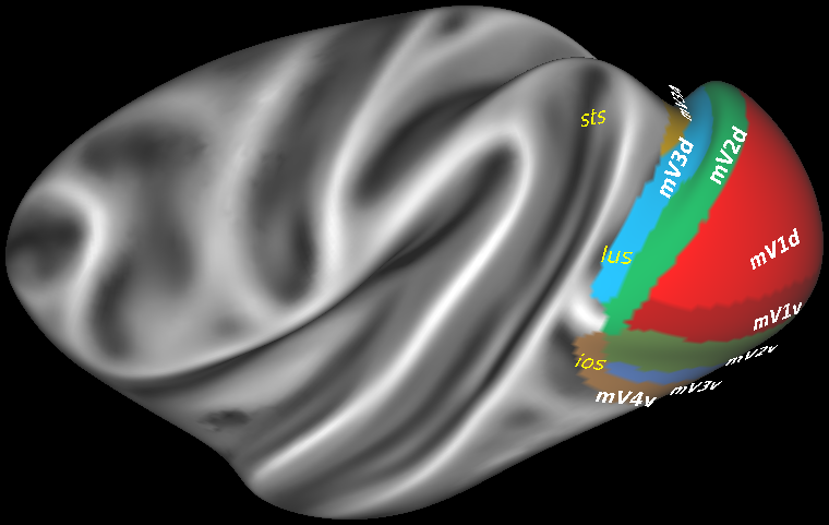
Parcellation scheme of the macaque early visual areas projected onto the lateral view of the Yerkes19 surface (Donahue et al., 2016). Eight cyto- and receptor architectonically distinc areas were ideintified in this brain region: dorsal and ventral subdivisions of the primary visual cortex (dV1 and vV1, respectively), area V2 (dV2 and vV2, respectively) and of area V3 (dV3 and vV3, respectively), as well as areas V3A dorsally and V4v ventrally.
ABSTRACT:
Existing cytoarchitectonic maps of the human and macaque posterior occipital cortex differ in the number of areas they display, thus hampering identification of homolog structures. We applied quantitative in vitro receptor autoradiography to characterize the receptor architecture of the primary visual and early extrastriate cortex in macaque and human brains, using previously published cytoarchitectonic criteria as starting point of our analysis. We identified 8 receptor architectonically distinct areas in the macaque brain (mV1d, mV1v, mV2d, mV2v, mV3d, mV3v, mV3A, mV4v), and their respective counterpart areas in the human brain (hV1d, hV1v, hV2d, hV2v, hV3d, hV3v, hV3A, hV4v). Mean densities of 14 neurotransmitter receptors were quantified in each area, and ensuing receptor fingerprints used for multivariate analyses. The 1st principal component segregated macaque and human early visual areas differ. However, the 2nd principal component showed that within each species, area-specific differences in receptor fingerprints were associated with the hierarchical processing level of each area. Subdivisions of V2 and V3 were found to cluster together in both species and were segregated from subdivisions of V1 and from V4v. Thus, comparative studies like this provide valuable architectonic insights into how differences in underlying microstructure impact evolutionary changes in functional processing of the primate brain and, at the same time, provide strong arguments for use of macaque monkey brain as a suitable animal model for translational studies.
PUBLICATION:
Brain Structure and Function
- DOI:
10.1007/s00429-021-02437-y
- Lucija Rapan
- Meiqi Niu
- Ling Zhao
- Thomas Funck
- Katrin Amunts
- Karl Zilles
- Nicola Palomero-Gallagher
- C. & O. Vogt Institute for Brain Research, Heinrich-Heine-University, 40225 Düsseldorf, Germany
- Department of Psychiatry, Psychotherapy, and Psychosomatics, Medical Faculty, RWTH Aachen
- Institute of Neuroscience and Medicine (INM-1), Research Centre Jülich, Jülich, Germany