


FULL TITLE:
Graded encoding of spatial novelty scales in the human brain
SPECIES:
Human
ABSTRACT:
Successful navigation relies on the ability to process and encode detailed information about our dynamic environments. Beyond familiarity, emerging studies now highlight the crucial role of novelty detection in this process, the precise neural mechanism of which remains poorly understood. Using ultra-high field 7T fMRI, we investigated how the human brain encodes spatial novelty during virtual navigation, with a particular focus on graded representations that follow systematic transitions between novel and familiar spaces. Our results revealed novelty and familiarity specific neural responses within the posterior and anterior poles of the bilateral hippocampus, respectively. On the cortical surface, two separable streams of activity patterns were observed in which regions within the visual and frontoparietal networks showed novelty-specific activity, while somatomotor, ventral attention and default mode regions preferred spatial familiarity. Importantly, we identified a distinct gradient along the hippocampal long axis and demonstrated the extended contribution of the posterior medial cortex to the encoding of spatial novelty scales that were intrinsically coupled with the hippocampal gradient. These findings advance our understanding of how the human brain encodes and processes spatial information, suggesting that graded representations of spatial novelty may serve as a fundamental organizational principle for spatial cognition in the human brain.
PUBLICATION:
Nature Communications
- Jörn A. Quent
- Liangyue Song
- Yueting Su
- Wenwen Yu
- He Wang
- Xinyu Liang
- Deniz Vatansever
- Fudan University
-
SpaNov_paper_revised.scene
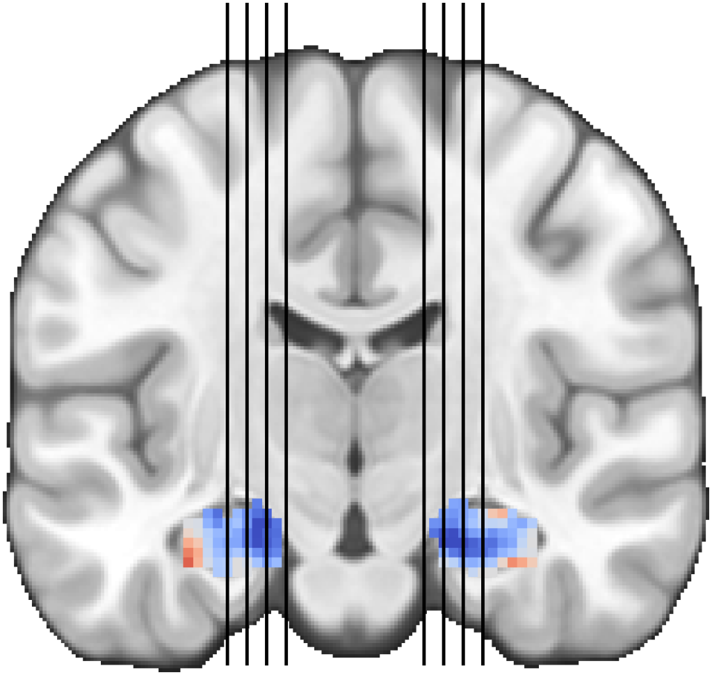
SCENES:- Figure 2a: Coronal slice showing position of sagittal slices
- Figure 2a: Grid of sagittal slices
- Figure 3a: Novelty familiarity contrast in cortex
- Figure 3b: Novelty/familiarity gradient along the posterior parietal cortex
- Supplementary map: Cluster IDs for novelty familiarity contrast
- Figure S4 b & c: Spatial novelty uncorrected vs. corrected for centrality
- Supplementary map: Correlation between spatial novelty contrast and centrality
- Figure S8b & c: Linear contrast across all object ranks (encoding success)