
Scene Preview
study:
Neurite imaging reveals microstructural variations in human cerebral cortical gray matter.
SCENE FILE:
Figures_NODDI
SCENE:
FigS4
DESCRIPTION:
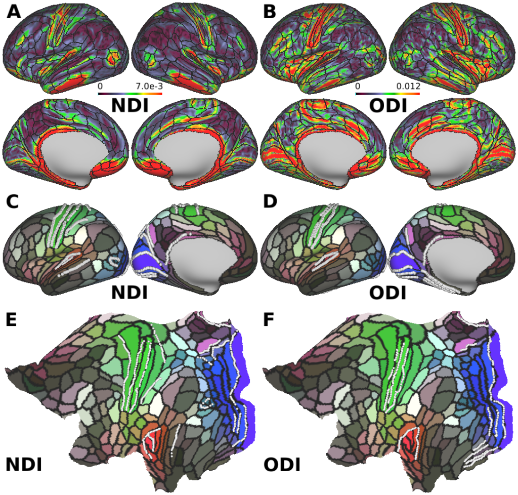
Comparison of 505-subject mean gradient maps and borders of neurite density index (NDI) and orientation dispersion index (ODI) with the Human Connectome Project (HCP) parcellations of the cerebral cortex (Glasser et al., 2016). (A) The gradient of NDI exhibits in strong gradient changes in several but not all borders of cortical areas, indicating that neurite density varies between these cortical parcels. (B) The gradient of ODI shows less correspondence to the borders, and high gradients are found within some of parcellations, such as primary visual, motor, somatosensory areas. The borders estimated from gradient of NDI (C,E, white dots) correspond well to the HCP parcellations (color label and black borders) shown in inflated surface (C) and flat map (E). The borders estimated from gradient of ODI (D,F white dots) show some correspondence with the HCP parcellations, but less than for NDI.
TAGS:
Surface Mesh:32k fs LR, Registration:MSMAll, Species:Human