
Scene Preview
study:
Human Habenula Functional Connectivity
SCENE FILE:
Ely_Hb_connectivity_supplementary
SCENE:
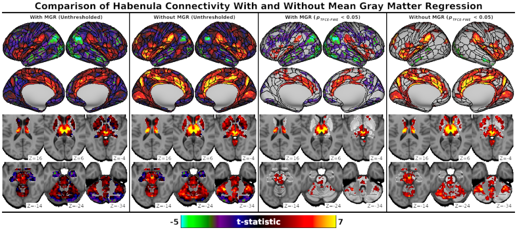
Fig. S3, Comparison of Habenula Connectivity With and Without Mean Gray Matter Regression
DESCRIPTION:
Fig. S3: Unthresholded (left) and significance-thresholded (pTFCE-FWE < 0.05, right) whole-brain Hb connectivity without (i.e., main analysis) and with mean gray matter timeseries regression (MGR). Unthresholded Hb connectivity appeared fairly similar in both analyses; as expected, MGR decreased the extent of positive connectivity and increased the extent of negative connectivity. Differences were more pronounced after significance thresholding. Following MGR, positive Hb connectivity with the VTA and Salience Network (e.g., dorsal ACC, anterior insula, supramarginal gyrus) was reduced but remained significant, while connectivity was no longer significant with early sensory areas (e.g., primary visual cortex). Significant negative Hb connectivity after MGR expanded to include the majority of the Default Mode Network (e.g., posterior cingulate, vmPFC, dmPFC, posterior lateral parietal cortex) as well as extrastriate visual and auditory association cortices, face and upper limb subareas of the somatomotor strip, and large portions of the lateral PFC.
TAGS:
Surface Mesh:32k fs LR, Registration:MSMAll, Atlas:Conte69, Species:Human