
Scene Preview
study:
The Impact of Traditional Neuroimaging Methods on the Spatial Localization of Cortical Areas
SCENE FILE:
Coalson_et_al_2018_suppl
SCENE:
Supplemental Figure S1
DESCRIPTION:
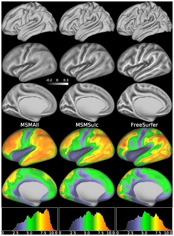
Top row shows the group average midthickness surfaces, where the columns indicate whether the results are from MSMAll, MSMSulc, or FreeSurfer registration, respectively. Rows 2 and 3 show the group average curvature maps. The bottom 3 rows show the cross-subject variability of the midthickness surface coordinates at each vertex (root mean square of the distance from the average coordinate - similar to standard deviation). Laterally, most of cortex is highly variable, with the exceptions in the central sulcus and the insula (where the curvature maps from all methods look similar), whereas medially, there is less variability overall. With folding-based registrations, the coordinate variability values are lower (FreeSurfer’s mean is 5.4 and MSMSulc’s mean is 5.5) than with areal-feature-based registrations (MSMAll’s mean is 6.2). This increased folding variability with areal-feature-based registration is another indication that cortical areas do not have consistent locations relative to folding patterns, which themselves are quite variable across subjects, even in tight folding registrations like FreeSurfer’s. Notably, FreeSurfer also has higher peaks of variability than MSMSulc, which was tuned to maximize functional alignment using folds and fits them less tightly than FreeSurfer (i.e. by constraining the registration to allow less distortion). This finding likely reflects overfitting of incompatible folding patterns by FreeSurfer registration. See Supplemental Methods Section M5.
TAGS:
Surface Mesh:32k fs LR, Registration:MSMAll, Registration:MSMSulc