
Scene Preview
study:
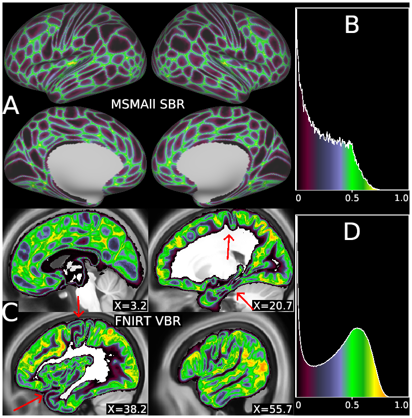
The Impact of Traditional Neuroimaging Methods on the Spatial Localization of Cortical Areas
SCENE FILE:
Coalson_et_al_2018
SCENE:
Figure 3
DESCRIPTION:
Areal uncertainty of MSMAll surface-based alignment (A) versus FNIRT volume-based alignment (C) for the 210V probabilistic cortical areas. The traditional volume-based approach has substantially greater uncertainty (greens, yellows, and oranges) than the HCP-style surface-based approach as seen in the histograms (B and D) as well as the images (A and C). In the volume-based results, some locations have low uncertainty (purple and black) and relatively sharp boundaries between areas (red arrows: early sensorimotor, insular, and inferior temporal cortex), comparable to what is consistently found on the surface. The volume ROIs that were used to create this figure were generated by mapping the individuals’ parcellations to the 0.7-mm MNI template space using the indiivduals’ native resolution MNI space surfaces and the ribbon-mapping method (19). Using 0.7-mm voxels minimizes the effects of voxel size on the group probability maps, allowing the effect of alignment to be investigated separately from the effect of voxel resolution. In practice, typical fMRI resolutions lead to increased signal mixing between areas and noncortical tissues, for both surface and volume analysis (see Effect of Acquisition Resolution). See SI Appendix, Supplemental Methods M2, M3, and M8.
TAGS:
Surface Mesh:32k fs LR, Registration:MSMAll, Modality:T1-weighted, Other Data:gene