
Scene Preview
study:
The Impact of Traditional Neuroimaging Methods on the Spatial Localization of Cortical Areas
SCENE FILE:
Coalson_et_al_2018_suppl
SCENE:
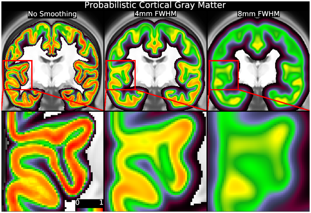
Supplemental Figure S7
DESCRIPTION:
This figure shows the biasing effects of volume-based smoothing on cortical geometry. Top row: Probabilistic maps of cortical gray matter for different smoothing levels shown in a coronal slice. Bottom row: Inset close-ups of a portion of the cortical probability at different smoothing levels. The white dots are at the same spatial coordinates in each image. Without smoothing, features such as the fundus of the circular sulcus (around the insula) and the superior temporal gyrus, are sharply defined, as is the boundary between gray and white matter in this region. With smoothing (particularly at 8 mm FWHM, right column), the sulcal fundi are not only blurry, but the apparent location of the transition between gray and white matter has substantially shifted. These biasing effects occur because cortical folding leads to more cortical voxels on one side of each gyral or sulcal curve. Clusters of nearby cortical voxels increase the smoothed values of the cortical probability in their vicinity, resulting in a bias of higher values towards one side of the original curved cortical ribbon. For cortical areas on gyral crowns or in sulcal fundi, or for groups of nearby areas that often activate together, the increased correlation with nearby cortical voxels will cause a similar volume-based smoothing bias towards the inside of the curvature. Thus, volume-based smoothing distorts important spatial relationships relevant to cortical functional organization. These effects further complicate efforts to map legacy volume-smoothed data onto the cortical surface (see Main Text Section "Mapping legacy group average volume results onto the surface"). See Supplemental Methods Sections M3 and M5.
TAGS:
Modality:T1-weighted