
Scene Preview
Scene: Figure_8
study:
Basis of executive functions
SCENE FILE:
ExecFunc_ms
SCENE:
Figure_8
DESCRIPTION:
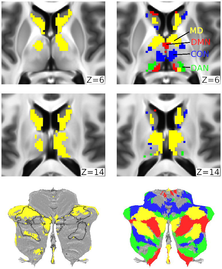
(a) Subcortical axial slices and a cerebellar flat map showing surviving voxels in caudate, thalamus and cerebellum for the conjunction of significantly activated voxels for each of the three executive contrasts (p<0.05 FDR corrected for each structure separately). (b) Voxels belonging to each network (yellow=MD, green=DAN, blue=CON, red=DMN). MD caudate, thalamic and cerebellar voxels are from (Assem et al. 2020). The other three RSN definitions are from (Ji et al. 2019).
TAGS:
Species:Human, Modality:Task fMRI, Modality:T1-weighted, Modality:T2-weighted