
Scene Preview
study:
Brain/MINDS Beyond Human Brain MRI Study: Multi-Site Harmonization
SCENE FILE:
Figures
SCENE:
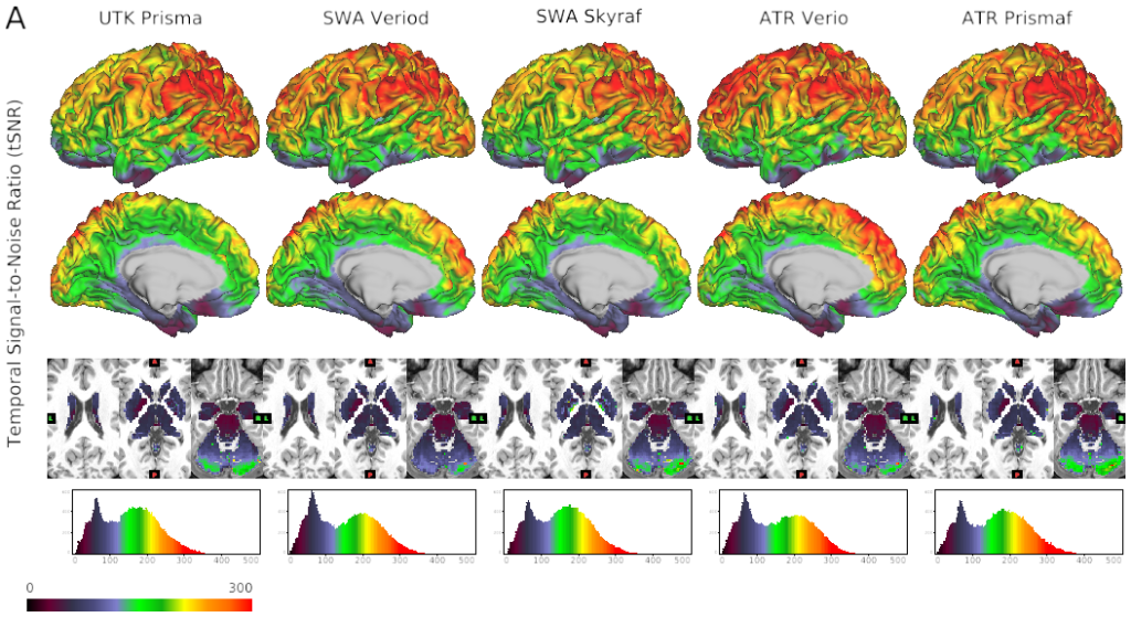
Figure 3A tSNR - fMRI
DESCRIPTION:
Quality of MRI and preliminary cortical structures obtained by HARP in a single traveling subject across scanners/sites.
A) Temporal signal-to-noise ratio (tSNR) obtained in a single subject (ID = 9503) across different scanners/sites by a harmonized MRI protocol (a sequence of functional MRI in HARP using a multi-band echo planar imaging with TR/TE = 800/34.4 ms; see Supplementary Table S1 for other details). The images from top to bottom show color-coded tSNR maps in 32k greyordinates (see main text) overlaid on the lateral and medial surface of the mid-thickness surface of the left hemisphere, the subcortical sections of the T1W image, and the histogram of the tSNR values.
TAGS:
Surface Mesh:32k fs LR, Registration:MSMAll, Modality:T1-weighted, Modality:T2-weighted