
Scene Preview
study:
Human Habenula Functional Connectivity
SCENE FILE:
Ely_Hb_connectivity
SCENE:
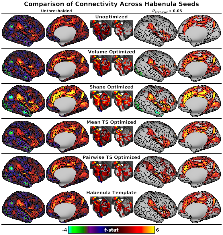
Fig. 3, Comparison of Connectivity Across Habenula Seeds
DESCRIPTION:
Fig. 3: Unthresholded (left) and significance-thresholded (pTFCE-FWE < 0.05, right) whole-brain connectivity results for the six types of Hb seed ROIs (see Fig. 2), displayed on the “very inflated” right cortical surface with areal boundary lines from the multimodal HCP parcellation (Glasser et al. 2016a) and a midsagittal slice through subcortical MNI space (X = 0). Unthresholded connectivity patterns were largely consistent across Hb seeds, including positive connectivity with the dorsal ACC, mid-cingulate, insula, posterior precuneus, and VTA. However, strength of connectivity with these areas varied between Hb seeds, resulting in larger apparent differences after significance thresholding (e.g., primary visual cortex, dlPFC). Connectivity was generally strongest for Shape Optimized ROIs and weakest for Unoptimized ROIs. Left hemisphere results (not presented) were very similar.
TAGS:
Surface Mesh:32k fs LR, Atlas:Conte69, Species:Human, Registration:MSMAll, Other Data:gene