
Scene Preview
study:
The Impact of Traditional Neuroimaging Methods on the Spatial Localization of Cortical Areas
SCENE FILE:
Coalson_et_al_2018
SCENE:
Figure 5
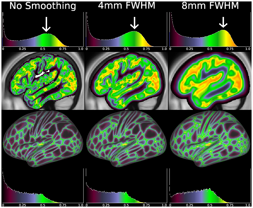
DESCRIPTION:
Comparison of different degrees of smoothing (columns) for both volume-based (Upper two rows) and surface-based (Lower two rows) approaches. Both areal uncertainty maps and histograms are shown. These were computed by smoothing the probability maps, which is equivalent to smoothing the per subject ROIs before averaging. Smoothing kernels on the surface clearly have less deleterious effects than smoothing kernels of the same size in the volume, because surface smoothing avoids smoothing across sulci or into other tissues. As with Fig. 3, the volume-based histograms have substantial “low uncertainty” tails that arise from poor alignment of the cortical ribbon, and from the tail of the Gaussian smoothing kernel within the white matter and CSF. See SI Appendix, Supplemental Methods M3, M4, and M8.
TAGS:
Surface Mesh:32k fs LR, Registration:MSMAll, Modality:T1-weighted