
Scene Preview
study:
The Impact of Traditional Neuroimaging Methods on the Spatial Localization of Cortical Areas
SCENE FILE:
Coalson_et_al_2018
SCENE:
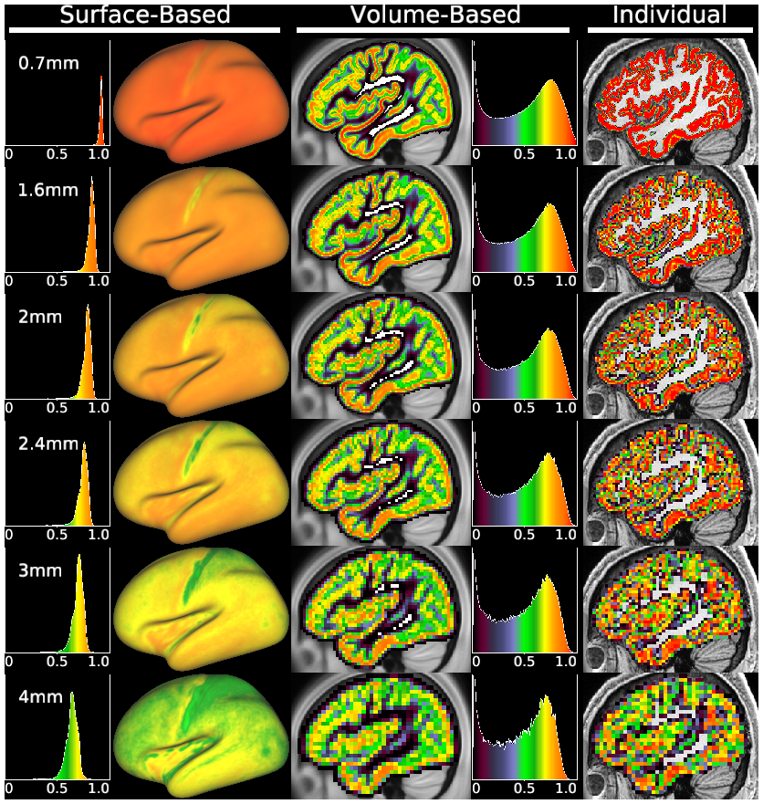
Figure 7
DESCRIPTION:
The effect of acquisition resolution on the separation of cortical signal from noncortical signal, for surface-based (Left two columns) and volume-based (Center two columns) processing. The measure shown is the group average cortical gray matter fraction of each vertex or voxel. The Right-most column shows an individual’s (HCP subject 121618) cortical fraction volumes for the same six resolutions, as an example of the inputs to the analyses. Smoothing was not applied. The cortical signal fraction becomes somewhat degraded at the edge of cortex (green voxels) in many regions, even at 2-mm resolution (even though this is less than the mean cortical thickness) and is severely degraded (many green and blue voxels) at traditionally used resolutions between 3 and 4 mm. See SI Appendix, Supplemental Methods M2.
TAGS:
Surface Mesh:32k fs LR, Registration:MSMAll, Modality:T1-weighted