
Scene Preview
study:
Human Habenula Functional Connectivity
SCENE FILE:
Ely_Hb_connectivity_supplementary
SCENE:
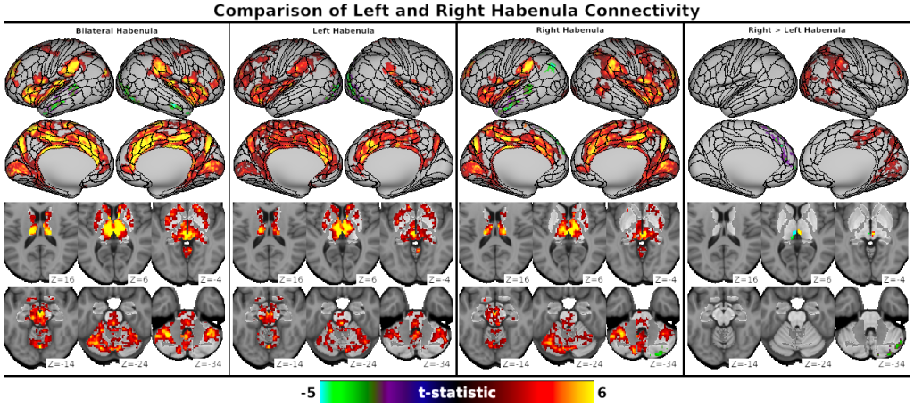
Fig. S2, Comparison of Left and Right Habenula Connectivity
DESCRIPTION:
Fig. S2: Whole-brain resting-state functional connectivity results for bilateral (i.e., main analysis), left, and right Shape Optimized seed ROIs and the contrast of left vs. right ROIs. Connectivity patterns were similar for left and right Hb ROIs and resembled bilateral Hb ROI connectivity. Connectivity was generally stronger for structures ipsilateral vs. contralateral to the unihemispheric Hb ROIs (e.g., insula, dlPFC). Directly contrasting left vs. right Hb connectivity (rightmost panel) using paired t-tests also indicated limited lateralization: right Hb connectivity (red/yellow) was significantly stronger with the right visual, mid-/posterior cingulate, and lateral parietal cortices, while left Hb connectivity (blue/green) was significantly stronger with the left dmPFC and right posterior cerebellum. Results are displayed at the pTFCE-FWE < 0.05 threshold.
TAGS:
Surface Mesh:32k fs LR, Registration:MSMAll, Atlas:Conte69, Species:Human, Other Data:gene