
Scene Preview
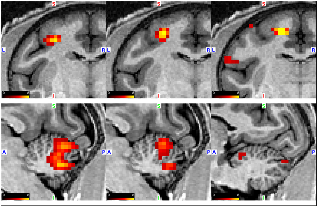
Scene: Fig. 2A: Somatotopic cerebellar activations evoked by optogenetic intracortical microstimulation of the primary motor cortex (monkey C)
SCENE FILE:
Figs_Goda_et_al_OptofMRINHP
SCENE:
Fig. 2A: Somatotopic cerebellar activations evoked by optogenetic intracortical microstimulation of the primary motor cortex (monkey C)
DESCRIPTION:
Z-statistic color maps (stimulation versus no-stimulation, P < 0.05, FWE-corrected for multiple comparisons) were overlaid onto coronal T1w images of the left M1 (upper row) and sagittal images of the right cerebellum (lower row) for monkey C. Left, middle, and right panels represent maps with the stimulations of the M1 distal forelimb, proximal forelimb, and hindlimb regions, respectively.
TAGS:
Species:Macaque, Atlas:Yerkes 19