
Scene Preview
Scene: Fig. 5, Panel B, Habenula Connectivity vs. Cortical Salience Network
study:
Human Habenula Functional Connectivity
SCENE FILE:
Ely_Hb_connectivity
SCENE:
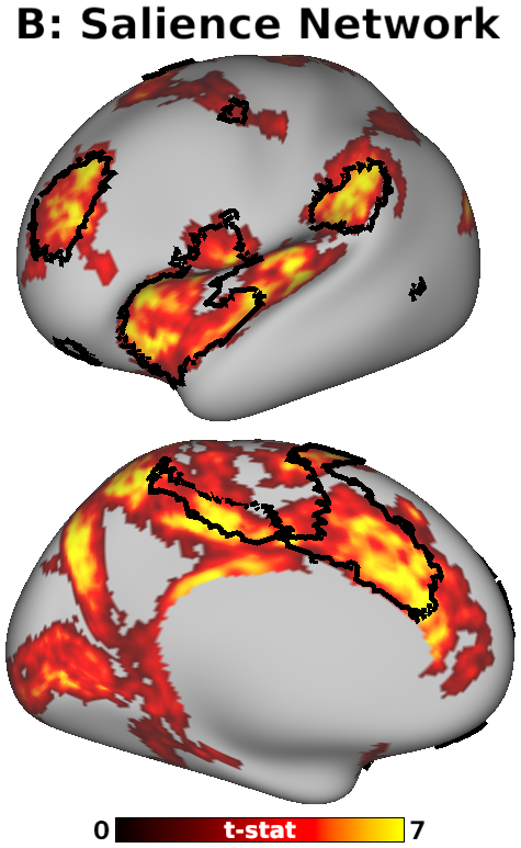
Fig. 5, Panel B, Habenula Connectivity vs. Cortical Salience Network
DESCRIPTION:
B: Significant (pTFCE-FWE < 0.05) positive Hb connectivity included nearly every part of the cortical Salience Network (outlined in black). Salience Network boundaries were created by combining networks 7 and 8 from the 17-network rs-fMRI parcellation published by Yeo et al. 2011 and distributed by the HCP. Displayed on the “very inflated” left cortical surface.
TAGS:
Surface Mesh:32k fs LR, Atlas:Conte69, Species:Human, Registration:MSMAll