
Scene Preview
study:
Sensory-biased lateral frontal cortex networks
SCENE FILE:
Tobyne2017_HCP_AV_LFC
SCENE:
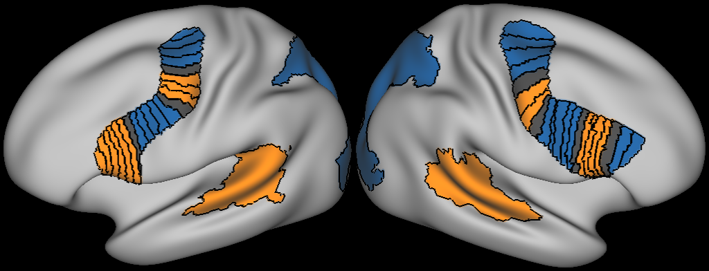
Figure 4: Lateral frontal hull rsFC analysis
DESCRIPTION:
Representation of the lateral frontal hull in each hemisphere. Each of the 21 segments are colored according to their bias (blue = visual, orange = auditory, gray = no bias)
Window 1:
Browser Tab 1:
Cerebral Montage:
Left Surface:
Surface: Q1-Q6_R440.L.inflated.32k_fs_LR.surf.gii
Structure: CortexLeft
Primary Type: Inflated
Secondary Type: Midthickness
Right Surface:
Surface: Q1-Q6_R440.R.inflated.32k_fs_LR.surf.gii
Structure: CortexRight
Primary Type: Inflated
Secondary Type: Midthickness
Selected Views: Lateral
Overlay Set
Overlay 1:
File: sliced_hull.fs_LR.dlabel.nii
Map Index: 1
Map Name: node label
Structure: All
Overlay 2:
File: prob_labels.fs_LR.dlabel.nii
Map Index: 1
Map Name: node label
Structure: All
TAGS:
Surface Mesh:32k fs LR, Species:Human